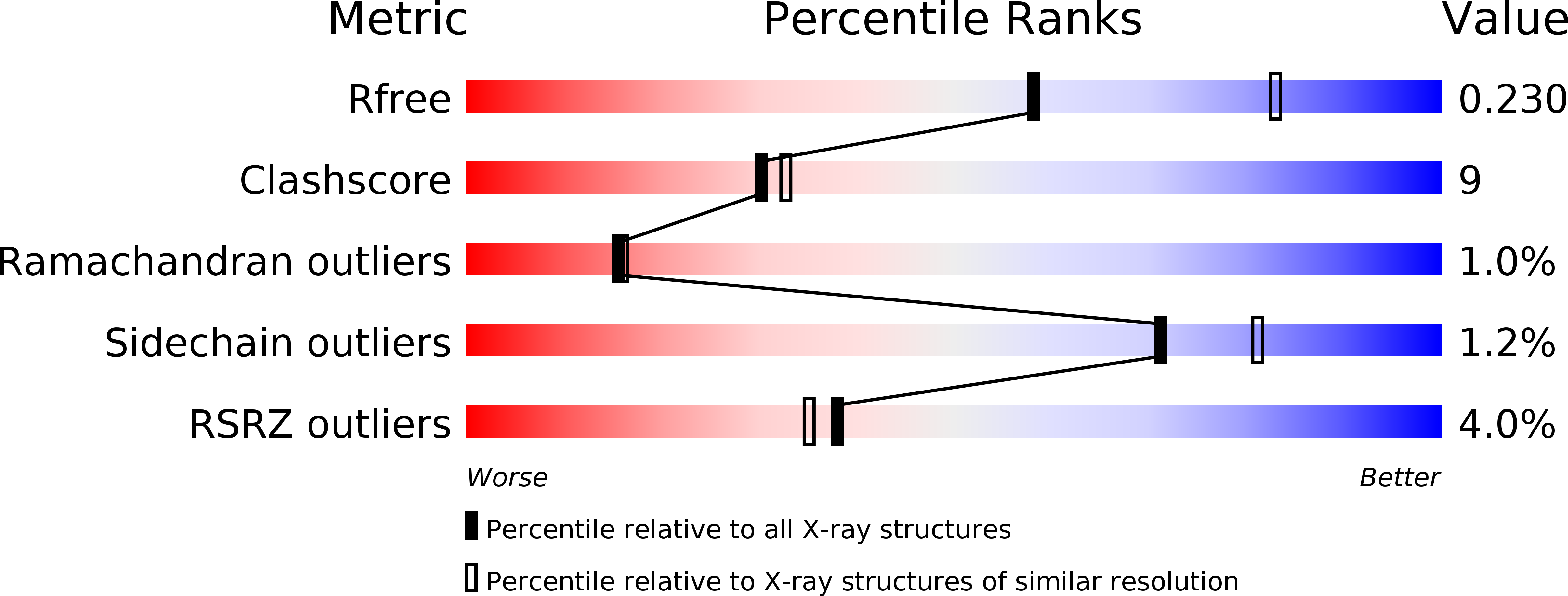
3E90 image
Deposition Date 2008-08-21
Release Date 2009-01-20
Last Version Date 2024-10-30
Entry Detail
PDB ID:
3E90
Keywords:
Title:
West Nile vi rus NS2B-NS3protease in complexed with inhibitor Naph-KKR-H
Biological Source:
Source Organism:
Host Organism:
Method Details:
Experimental Method:
Resolution:
2.45 Å
R-Value Free:
0.24
R-Value Work:
0.19
Space Group:
C 2 2 21
Macromolecular Entities
Structures with similar UniProt ID
Protein Blast
Polymer Type:polypeptide(L)
Molecule:NS2B cofactor
Chain IDs:A, C
Chain Length:50
Number of Molecules:2
Biological Source:West Nile virus
Structures with similar UniProt ID
Protein Blast
Polymer Type:polypeptide(L)
Molecule:NS3 protease
Chain IDs:B, D
Chain Length:198
Number of Molecules:2
Biological Source:West Nile virus
Ligand Molecules
Primary Citation
Structure of West Nile virus NS3 protease: ligand stabilization of the catalytic conformation
J.Mol.Biol. 385 1568 1577 (2009)
PMID: 19059417 DOI: 10.1016/j.jmb.2008.11.026

Abstact

Over the last decade, West Nile virus has spread rapidly via mosquito transmission from infected migratory birds to humans. One potential therapeutic approach to treating infection is to inhibit the virally encoded serine protease that is essential for viral replication. Here we report the crystal structure of the viral NS3 protease tethered to its essential NS2B cofactor and bound to a potent substrate-based tripeptide inhibitor, 2-naphthoyl-Lys-Lys-Arg-H (K(i)=41 nM), capped at the N-terminus by 2-naphthoyl and capped at the C-terminus by aldehyde. An important and unexpected feature of this structure is the presence of two conformations of the catalytic histidine suggesting a role for ligand stabilization of the catalytically competent His conformation. Analysis of other West Nile virus NS3 protease structures and related serine proteases supports this hypothesis, suggesting that the common catalytic mechanism involves an induced-fit mechanism.

Legend

Protein

Chemical

Disease

Primary Citation of related structures