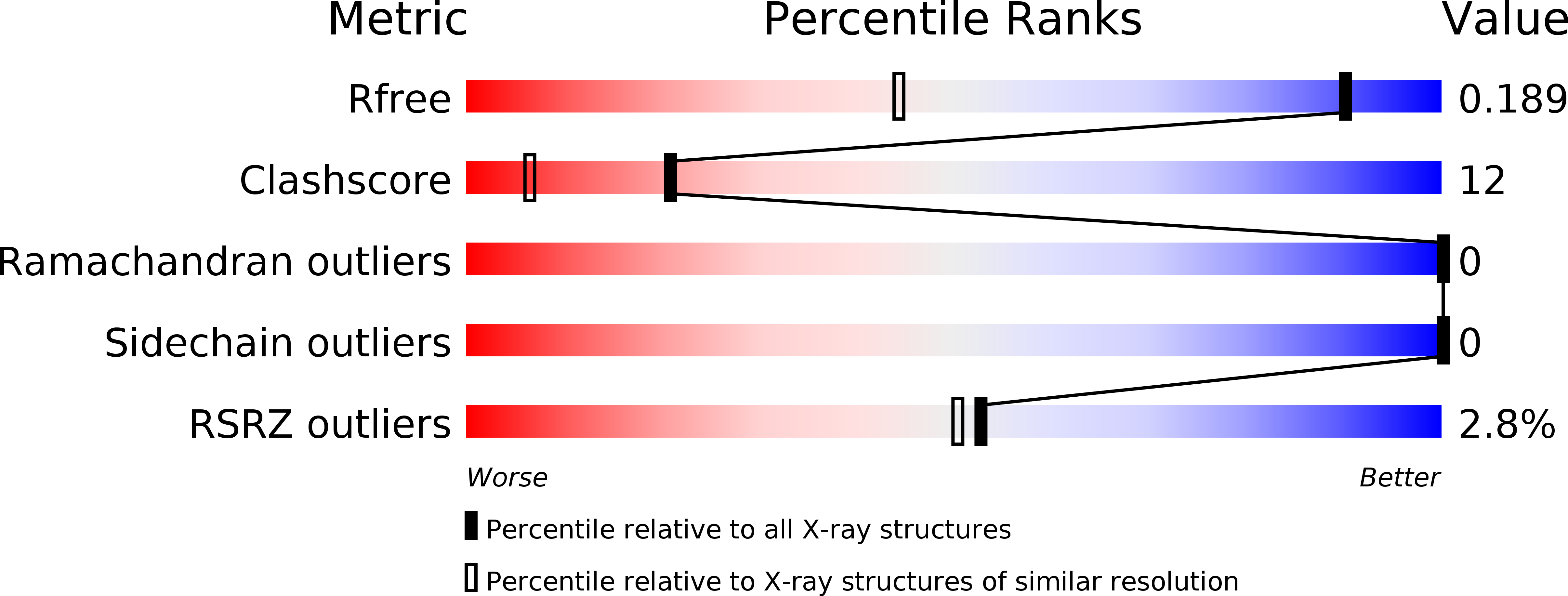
3E8T image
Deposition Date 2008-08-20
Release Date 2008-12-09
Last Version Date 2024-10-09
Entry Detail
PDB ID:
3E8T
Title:
Crystal Structure of Epiphyas postvittana Takeout 1
Biological Source:
Source Organism:
Host Organism:
Method Details:
Experimental Method:
Resolution:
1.30 Å
R-Value Free:
0.18
R-Value Work:
0.14
R-Value Observed:
0.14
Space Group:
P 1 21 1
Macromolecular Entities
Polymer Type:polypeptide(L)
Molecule:Takeout-like protein 1
Chain IDs:A
Chain Length:220
Number of Molecules:1
Biological Source:Epiphyas postvittana
Ligand Molecules
Primary Citation
Crystal structure of Epiphyas postvittana takeout 1 with bound ubiquinone supports a role as ligand carriers for takeout proteins in insects
J.Biol.Chem. 284 3496 3503 (2009)
PMID: 19073605 DOI: 10.1074/jbc.M807467200

Abstact

Takeout (To) proteins are found exclusively in insects and have been proposed to have important roles in various aspects of their physiology and behavior. Limited sequence similarity with juvenile hormone-binding proteins (JHBPs), which specifically bind and transport juvenile hormones in Lepidoptera, suggested a role for To proteins in binding hydrophobic ligands. We present the first crystal structure of a To protein, EpTo1 from the light brown apple moth Epiphyas postvittana, solved in-house by the single-wavelength anomalous diffraction technique using sulfur anomalous dispersion, and refined to 1.3 angstroms resolution. EpTo1 adopts the unusual alpha/beta-wrap fold, seen only for JHBP and several mammalian lipid carrier proteins, a scaffold tailored for the binding and/or transport of hydrophobic ligands. EpTo1 has a 45 angstroms long, purely hydrophobic, internal tunnel that extends for the full length of the protein and accommodates a bound ligand. The latter was shown by mass spectrometry to be ubiquinone-8 and is probably derived from Escherichia coli. The structure provides the first direct experimental evidence that To proteins are ligand carriers; gives insights into the nature of endogenous ligand(s) of EpTo1; shows, by comparison with JHBP, a basis for different ligand specificities; and suggests a mechanism for the binding/release of ligands.

Legend

Protein

Chemical

Disease

Primary Citation of related structures