3E4Z image
Deposition Date 2008-08-12
Release Date 2009-08-18
Last Version Date 2024-02-21
Entry Detail
PDB ID:
3E4Z
Title:
Crystal structure of human insulin degrading enzyme in complex with insulin-like growth factor II
Biological Source:
Source Organism:
Homo sapiens (Taxon ID: 9606)
Host Organism:
Method Details:
Experimental Method:
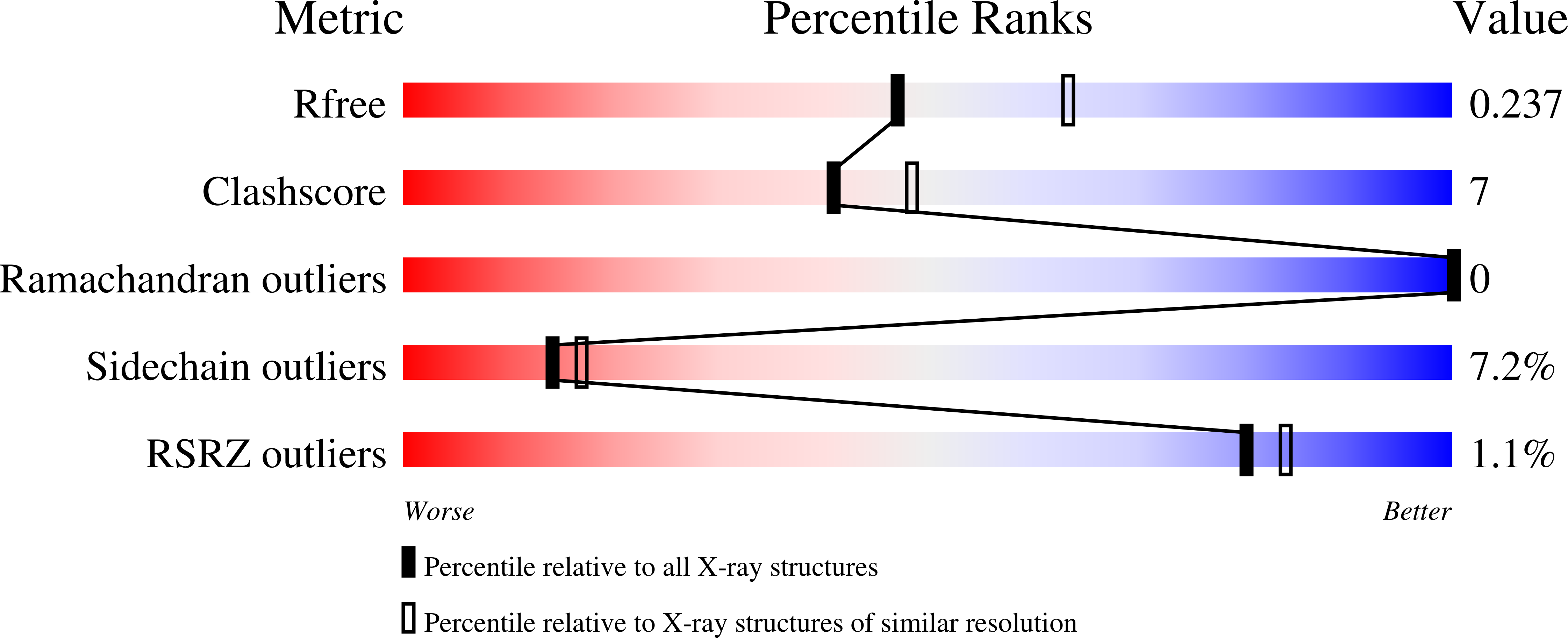
Resolution:
2.28 Å
R-Value Free:
0.22
R-Value Work:
0.19
R-Value Observed:
0.19
Space Group:
P 65
Macromolecular Entities
Structures with similar UniProt ID
Protein Blast
Polymer Type:polypeptide(L)
Molecule:Insulin-degrading enzyme
Mutagens:C110L, E111Q, C171S, C178A, C257V, C414L, C573N, C590S, C789S, C812A, C819A, C904S, W908Y
Chain IDs:A, B
Chain Length:990
Number of Molecules:2
Biological Source:Homo sapiens
Structures with similar UniProt ID
Protein Blast
Polymer Type:polypeptide(L)
Molecule:Insulin-like growth factor II
Gene (Uniprot):IGF2
Chain IDs:C, D
Chain Length:67
Number of Molecules:2
Biological Source:Homo sapiens
Ligand Molecules
Primary Citation
Molecular Basis for the Recognition and Cleavages of IGF-II, TGF-alpha, and Amylin by Human Insulin-Degrading Enzyme.
J.Mol.Biol. 395 430 443 (2010)
PMID: 19896952 DOI: 10.1016/j.jmb.2009.10.072

Abstact

Insulin-degrading enzyme (IDE) is involved in the clearance of many bioactive peptide substrates, including insulin and amyloid-beta, peptides vital to the development of diabetes and Alzheimer's disease, respectively. IDE can also rapidly degrade hormones that are held together by intramolecular disulfide bond(s) without their reduction. Furthermore, IDE exhibits a remarkable ability to preferentially degrade structurally similar peptides such as the selective degradation of insulin-like growth factor (IGF)-II and transforming growth factor-alpha (TGF-alpha) over IGF-I and epidermal growth factor, respectively. Here, we used high-accuracy mass spectrometry to identify the cleavage sites of human IGF-II, TGF-alpha, amylin, reduced amylin, and amyloid-beta by human IDE. We also determined the structures of human IDE-IGF-II and IDE-TGF-alpha at 2.3 A and IDE-amylin at 2.9 A. We found that IDE cleaves its substrates at multiple sites in a biased stochastic manner. Furthermore, the presence of a disulfide bond in amylin allows IDE to cut at an additional site in the middle of the peptide (amino acids 18-19). Our amylin-bound IDE structure offers insight into how the structural constraint from a disulfide bond in amylin can alter IDE cleavage sites. Together with NMR structures of amylin and the IGF and epidermal growth factor families, our work also reveals the structural basis of how the high dipole moment of substrates complements the charge distribution of the IDE catalytic chamber for the substrate selectivity. In addition, we show how the ability of substrates to properly anchor their N-terminus to the exosite of IDE and undergo a conformational switch upon binding to the catalytic chamber of IDE can also contribute to the selective degradation of structurally related growth factors.

Legend

Protein

Chemical

Disease

Primary Citation of related structures