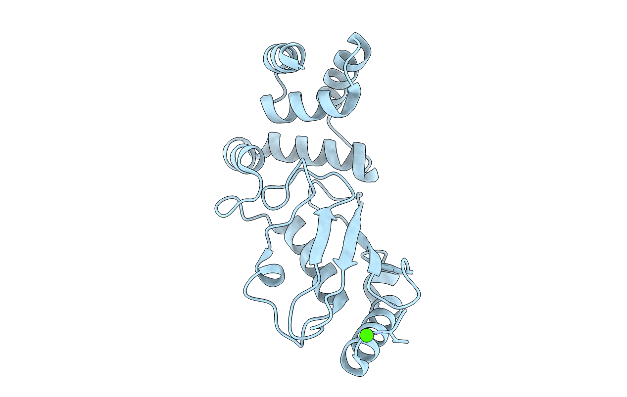
3E46 image
Entry Detail
PDB ID:
3E46
Keywords:
Title:
Crystal structure of ubiquitin-conjugating enzyme E2-25kDa (Huntington interacting protein 2) M172A mutant
Biological Source:
Source Organism:
Host Organism:
PDB Version:
Deposition Date:
2008-08-09
Release Date:
2008-08-26
Method Details:
Experimental Method:
Resolution:
1.86 Å
R-Value Free:
0.21
R-Value Work:
0.17
R-Value Observed:
0.17
Space Group:
I 4
Macromolecular Entities
Polymer Type:polypeptide(L)
Description:Ubiquitin-conjugating enzyme E2-25 kDa
Mutations:M172A
Chain IDs:A
Chain Length:253
Number of Molecules:1
Biological Source:Homo sapiens
Ligand Molecules
Primary Citation
Structure of full-length ubiquitin-conjugating enzyme E2-25K (huntingtin-interacting protein 2).
Acta Crystallogr.,Sect.F 65 440 444 (2009)
PMID: 19407372 DOI: 10.1107/S1744309109011117

Abstact

The ubiquitin-conjugating enzyme E2-25K has been identified as a huntingtin (the key protein in Huntington's disease) interacting protein and has been shown to play a role in mediating the toxicity of Abeta, the principal protein involved in Alzheimer's disease pathogenesis. E2-25K is a dual-domain protein with an ubiquitin-associated (UBA) domain as well as a conserved ubiquitin-conjugating (UBC) domain which catalyzes the formation of a covalent bond between the C-terminal glycine of an ubiquitin molecule and the -amine of a lysine residue on the acceptor protein as part of the ubiquitin-proteasome pathway. The crystal structures of E2-25K M172A mutant protein at pH 6.5 and pH 8.5 were determined to 1.9 and 2.2 A resolution, respectively. Examination of the structures revealed domain-domain interactions between the UBC and UBA domains which have not previously been reported.

Legend

Protein

Chemical

Disease

Primary Citation of related structures