3E3U image
Deposition Date 2008-08-08
Release Date 2009-01-20
Last Version Date 2024-02-21
Entry Detail
PDB ID:
3E3U
Keywords:
Title:
Crystal structure of Mycobacterium tuberculosis peptide deformylase in complex with inhibitor
Biological Source:
Source Organism:
Host Organism:
Method Details:
Experimental Method:
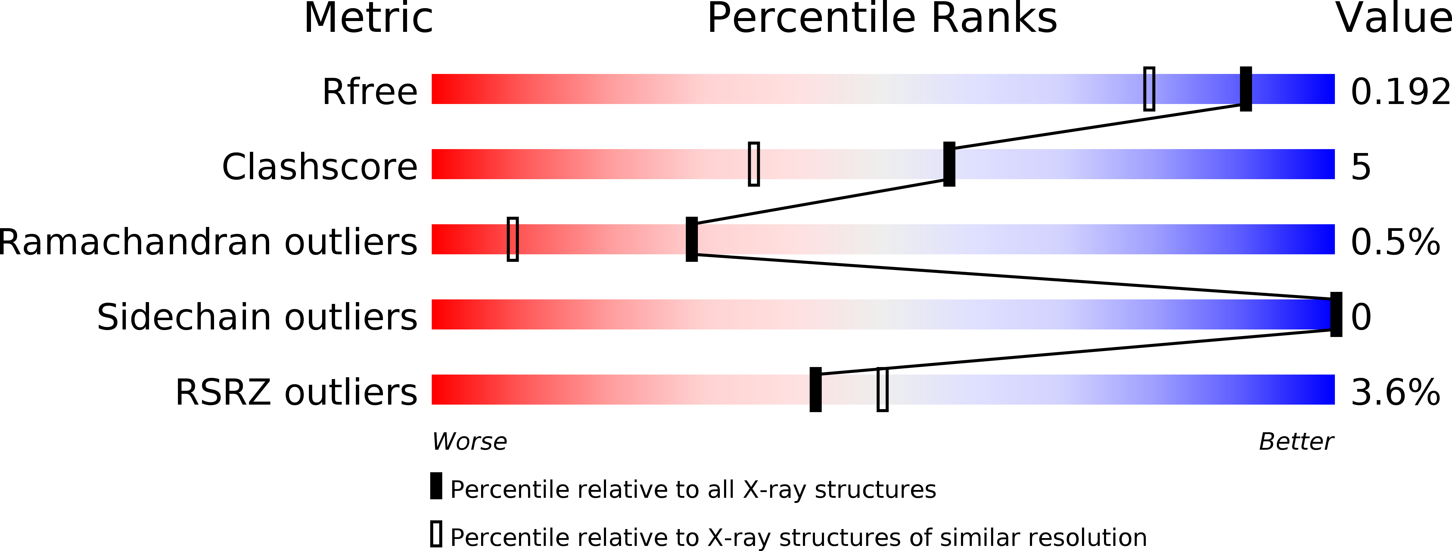
Resolution:
1.56 Å
R-Value Free:
0.20
R-Value Work:
0.17
R-Value Observed:
0.17
Space Group:
C 1 2 1
Macromolecular Entities
Polymer Type:polypeptide(L)
Molecule:Peptide deformylase
Chain IDs:A
Chain Length:197
Number of Molecules:1
Biological Source:Mycobacterium tuberculosis
Primary Citation
Peptide deformylase inhibitors of Mycobacterium tuberculosis: synthesis, structural investigations, and biological results.
Bioorg.Med.Chem.Lett. 18 6568 6572 (2008)
PMID: 19008098 DOI: 10.1016/j.bmcl.2008.10.040

Abstact

Bacterial peptide deformylase (PDF) belongs to a subfamily of metalloproteases catalyzing the removal of the N-terminal formyl group from newly synthesized proteins. We report the synthesis and biological activity of highly potent inhibitors of Mycobacterium tuberculosis (Mtb) PDF enzyme as well as the first X-ray crystal structure of Mtb PDF. Structure-activity relationship and crystallographic data clarified the structural requirements for high enzyme potency and cell based potency. Activities against single and multi-drug-resistant Mtb strains are also reported.

Legend

Protein

Chemical

Disease

Primary Citation of related structures