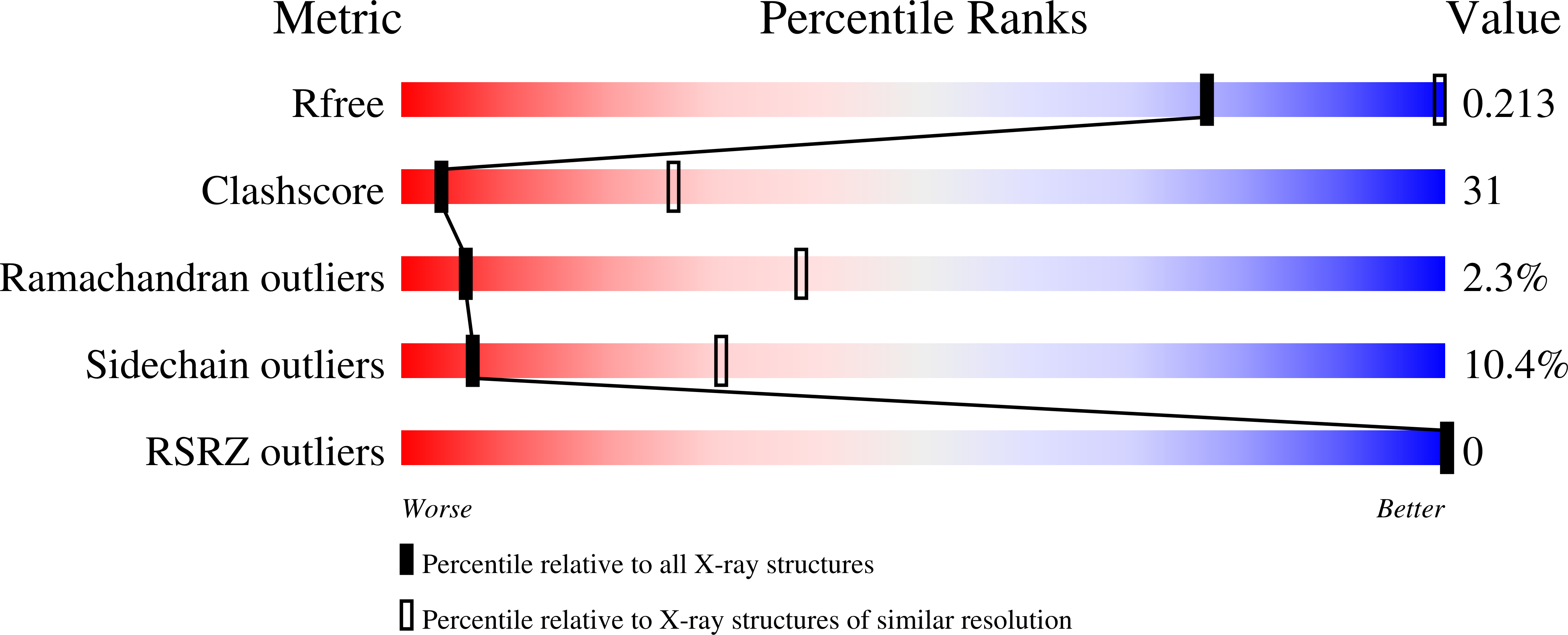
3E2H image
Deposition Date 2008-08-05
Release Date 2008-11-04
Last Version Date 2024-10-16
Entry Detail
PDB ID:
3E2H
Keywords:
Title:
Structure of the m67 high-affinity mutant of the 2C TCR in complex with Ld/QL9
Biological Source:
Source Organism:
Mus musculus (Taxon ID: 10090)
(Taxon ID: )
Host Organism:
Method Details:
Experimental Method:
Resolution:
3.80 Å
R-Value Free:
0.27
R-Value Work:
0.22
Space Group:
P 64 2 2
Macromolecular Entities
Polymer Type:polypeptide(L)
Molecule:H-2 class I histocompatibility antigen, L-D alpha chain
Gene (Uniprot):H2-L
Mutations:F8Y, V12T, P15R, I23T, N30D, A49V, I66V, R97W, K131R
Chain IDs:A
Chain Length:175
Number of Molecules:1
Biological Source:Mus musculus
Polymer Type:polypeptide(L)
Molecule:T-cell receptor alpha chain V region PHDS58
Mutations:L43P, W82R, G99L, F100E, A101R, S102P, A103Y
Chain IDs:C (auth: B)
Chain Length:109
Number of Molecules:1
Biological Source:Mus musculus
Polymer Type:polypeptide(L)
Molecule:M67 TCR beta chain
Chain IDs:D (auth: C)
Chain Length:110
Number of Molecules:1
Biological Source:Mus musculus
Polymer Type:polypeptide(L)
Molecule:QL9 PEPTIDE
Chain IDs:B (auth: Q)
Chain Length:9
Number of Molecules:1
Biological Source:
Ligand Molecules
Primary Citation
Distinct CDR3 Conformations in TCRs Determine the Level of Cross-Reactivity for Diverse Antigens, but Not the Docking Orientation
J.Immunol. 181 6255 6264 (2008)
PMID: 18941216

Abstact

T cells are known to cross-react with diverse peptide MHC Ags through their alphabeta TCR. To explore the basis of such cross-reactivity, we examined the 2C TCR that recognizes two structurally distinct ligands, SIY-K(b) and alloantigen QL9-L(d). In this study we characterized the cross-reactivity of several high-affinity 2C TCR variants that contained mutations only in the CDR3alpha loop. Two of the TCR lost their ability to cross-react with the reciprocal ligand (SIY-K(b)), whereas another TCR (m67) maintained reactivity with both ligands. Crystal structures of four of the TCRs in complex with QL9-L(d) showed that CDR1, CDR2, and CDR3beta conformations and docking orientations were remarkably similar. Although the CDR3alpha loop of TCR m67 conferred a 2000-fold higher affinity for SIY-K(b), the TCR maintained the same docking angle on QL9-L(d) as the 2C TCR. Thus, CDR3alpha dictated the affinity and level of cross-reactivity, yet it did so without affecting the conserved docking orientation.

Legend

Protein

Chemical

Disease

Primary Citation of related structures