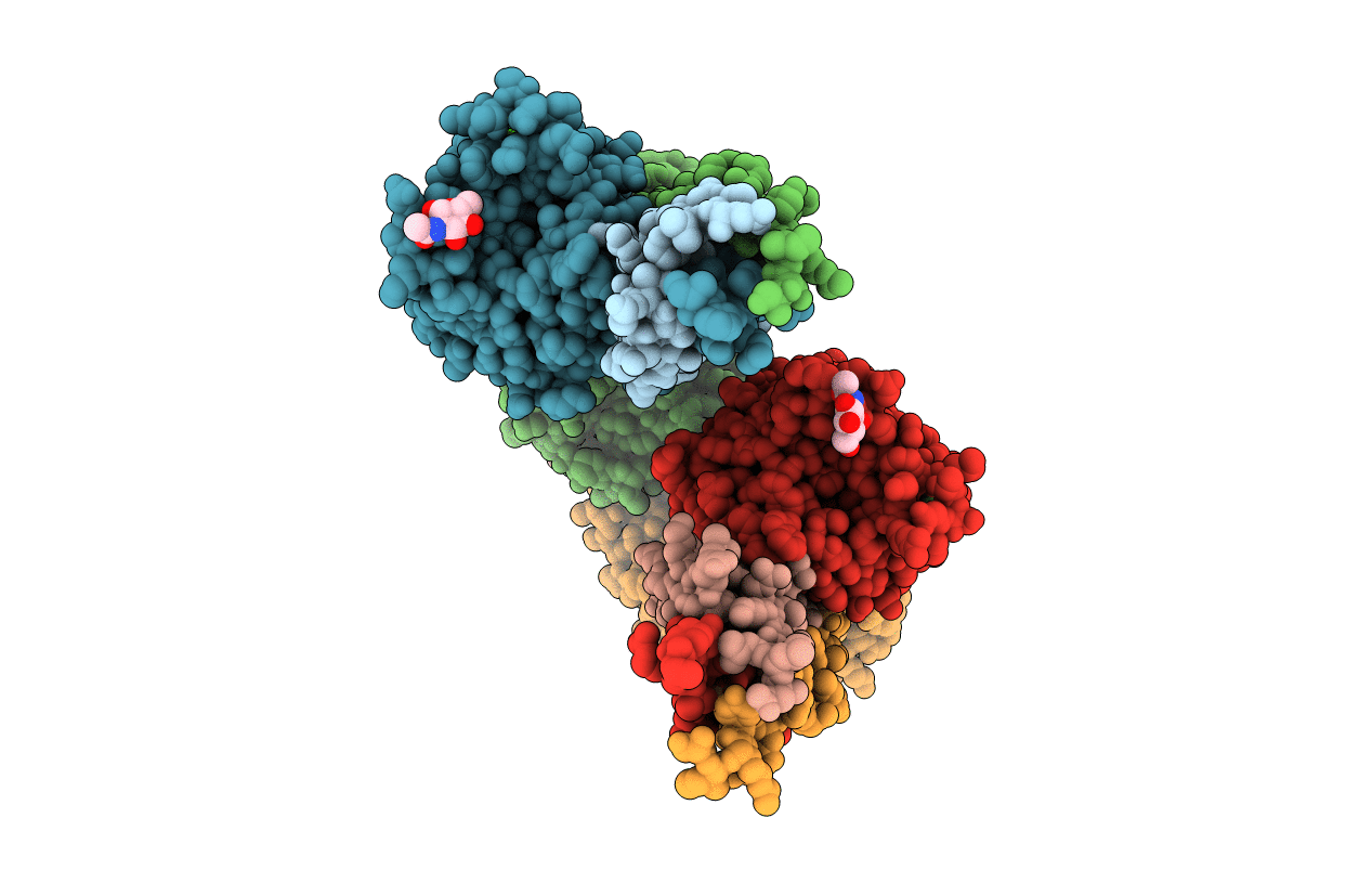
3E1I image
Deposition Date 2008-08-04
Release Date 2009-01-13
Last Version Date 2024-11-20
Entry Detail
PDB ID:
3E1I
Keywords:
Title:
Crystal Structure of BbetaD432A Variant Fibrinogen Fragment D with the Peptide Ligand Gly-His-Arg-Pro-amide
Biological Source:
Source Organism:
Host Organism:
Method Details:
Experimental Method:
Resolution:
2.30 Å
R-Value Free:
0.24
R-Value Work:
0.21
R-Value Observed:
0.21
Space Group:
P 21 21 21
Macromolecular Entities
Polymer Type:polypeptide(L)
Molecule:Fibrinogen alpha chain
Gene (Uniprot):FGA
Chain IDs:A, D
Chain Length:87
Number of Molecules:2
Biological Source:Homo sapiens
Polymer Type:polypeptide(L)
Molecule:Fibrinogen beta chain
Gene (Uniprot):FGB
Mutations:D432A
Chain IDs:B, E
Chain Length:328
Number of Molecules:2
Biological Source:Homo sapiens
Polymer Type:polypeptide(L)
Molecule:Fibrinogen gamma chain
Gene (Uniprot):FGG
Chain IDs:C, F
Chain Length:319
Number of Molecules:2
Biological Source:Homo sapiens
Polymer Type:polypeptide(L)
Molecule:Gly-His-Arg-Pro-amide
Chain IDs:G, H
Chain Length:5
Number of Molecules:2
Biological Source:synthetic construct
Primary Citation
Fibrinogen variant BbetaD432A has normal polymerization but does not bind knob "B".
Blood 113 4425 4430 (2009)
PMID: 19075185 DOI: 10.1182/blood-2008-09-178178

Abstact

Fibrinogen residue Bbeta432Asp is part of hole "b" that interacts with knob "B," whose sequence starts with Gly-His-Arg-Pro-amide (GHRP). Because previous studies showed BbetaD432A has normal polymerization, we hypothesized that Bbeta432Asp is not critical for knob "B" binding and that new knob-hole interactions would compensate for the loss of this Asp residue. To test this hypothesis, we solved the crystal structure of fragment D from BbetaD432A. Surprisingly, the structure (rfD-BbetaD432A+GH) showed the peptide GHRP was not bound to hole "b." We then re-evaluated the polymerization of this variant by examining clot turbidity, clot structure, and the rate of FXIIIa cross-linking. The turbidity and the rate of gamma-gamma dimer formation for BbetaD432A were indistinguishable compared with normal fibrinogen. Scanning electron microscopy showed no significant differences between the clots of BbetaD432A and normal, but the thrombin-derived clots had thicker fibers than clots obtained from batroxobin, suggesting that cleavage of FpB is more important than "B:b" interactions. We conclude that hole "b" and "B:b" knob-hole binding per se have no influence on fibrin polymerization.

Legend

Protein

Chemical

Disease

Primary Citation of related structures