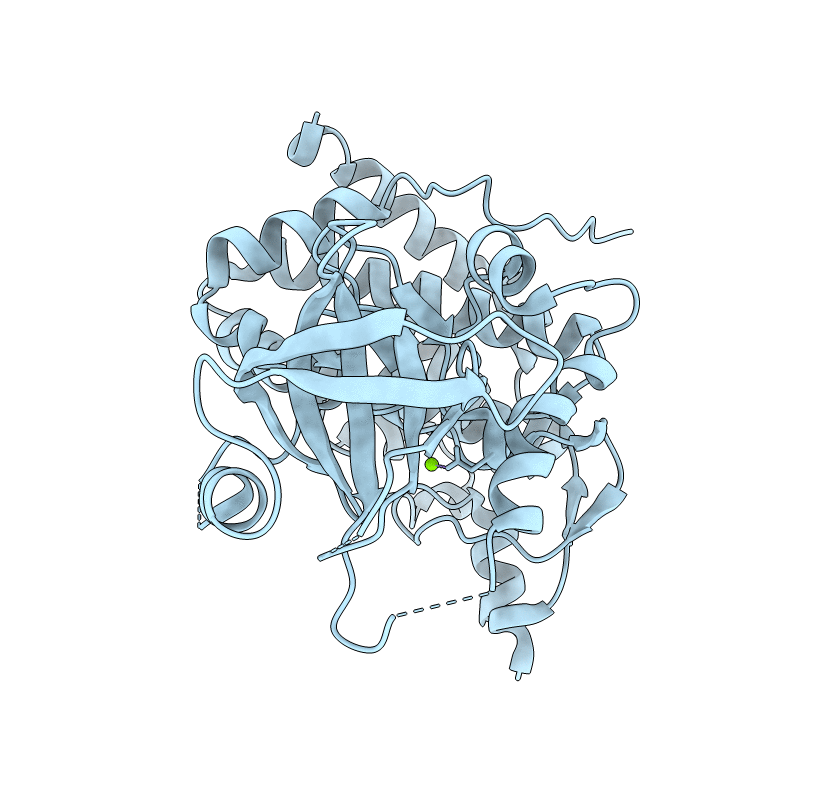
3DZO image
Deposition Date 2008-07-30
Release Date 2008-09-16
Last Version Date 2024-11-13
Entry Detail
PDB ID:
3DZO
Keywords:
Title:
Crystal structure of a rhoptry kinase from toxoplasma gondii
Biological Source:
Source Organism:
Host Organism:
Method Details:
Experimental Method:
Resolution:
1.80 Å
R-Value Free:
0.27
R-Value Work:
0.24
R-Value Observed:
0.24
Space Group:
P 1 21 1
Macromolecular Entities
Structures with similar UniProt ID
Protein Blast
Polymer Type:polypeptide(L)
Molecule:Rhoptry kinase domain
Chain IDs:A
Chain Length:413
Number of Molecules:1
Biological Source:Toxoplasma gondii
Ligand Molecules
Primary Citation
Novel structural and regulatory features of rhoptry secretory kinases in Toxoplasma gondii.
Embo J. 28 969 979 (2009)
PMID: 19197235 DOI: 10.1038/emboj.2009.24

Abstact

Serine/threonine kinases secreted from rhoptry organelles constitute important virulence factors of Toxoplasma gondii. Rhoptry kinases are highly divergent and their structures and regulatory mechanism are hitherto unknown. Here, we report the X-ray crystal structures of two related pseudokinases named ROP2 and ROP8, which differ primarily in their substrate-binding site. ROP kinases contain a typical bilobate kinase fold and a novel N-terminal extension that both stabilizes the N-lobe and provides a unique means of regulation. Although ROP2 and ROP8 were catalytically inactive, they provided a template for homology modelling of the active kinase ROP18, a major virulence determinant of T. gondii. Autophosphorylation of key residues in the N-terminal extension resulted in ROP18 activation, which in turn phosphorylated ROP2 and ROP8. Mutagenesis and mass spectrometry experiments revealed that ROP18 was maximally activated when this phosphorylated N-terminus relieved autoinhibition resulting from extension of aliphatic side chains into the ATP-binding pocket. This novel means of regulation governs ROP kinases implicated in parasite virulence.

Legend

Protein

Chemical

Disease

Primary Citation of related structures