Deposition Date
2008-07-28
Release Date
2009-01-27
Last Version Date
2024-10-09
Entry Detail
PDB ID:
3DYR
Keywords:
Title:
Crystal structure of E. coli thioredoxin mutant I76T in its oxidized form
Biological Source:
Source Organism(s):
Escherichia coli (Taxon ID: 83333)
Expression System(s):
Method Details:
Experimental Method:
Resolution:
2.00 Å
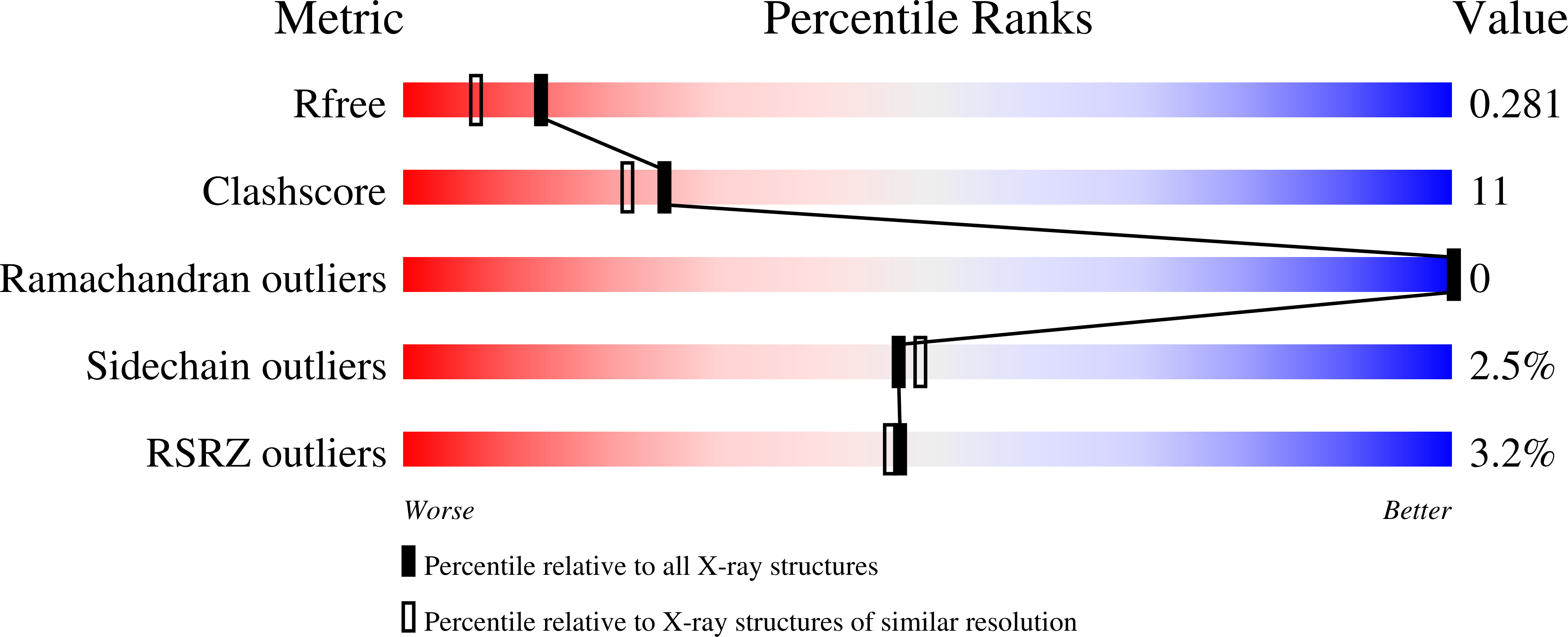
R-Value Free:
0.28
R-Value Work:
0.25
R-Value Observed:
0.25
Space Group:
C 1 2 1