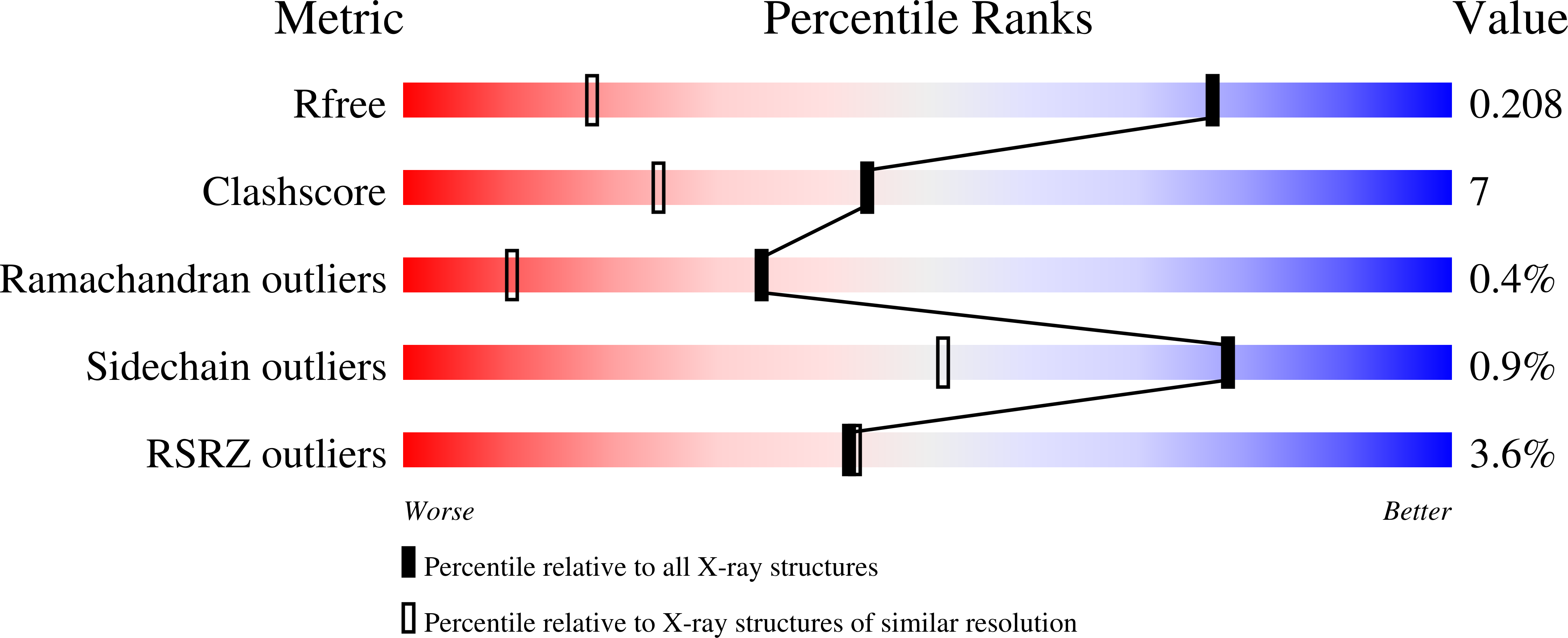
3DYB image
Deposition Date 2008-07-25
Release Date 2008-10-07
Last Version Date 2024-11-13
Entry Detail
PDB ID:
3DYB
Keywords:
Title:
proteinase K- digalacturonic acid complex
Biological Source:
Source Organism:
Method Details:
Experimental Method:
Resolution:
1.32 Å
R-Value Free:
0.20
R-Value Work:
0.15
R-Value Observed:
0.16
Space Group:
P 43 21 2
Macromolecular Entities
Structures with similar UniProt ID
Protein Blast
Polymer Type:polypeptide(L)
Molecule:Proteinase K
Gene (Uniprot):PROK
Chain IDs:A
Chain Length:279
Number of Molecules:1
Biological Source:Engyodontium album
Primary Citation
High-resolution structure of proteinase K cocrystallized with digalacturonic acid.
Acta Crystallogr.,Sect.F 65 192 198 (2009)
PMID: 19255463 DOI: 10.1107/S1744309109002218

Abstact

Proteinase K, a subtilisin-like fungal protease, was crystallized from a cocktail of small molecules containing digalacturonic acid (DGA). The crystal structure was determined to 1.32 A resolution and refined to an R factor of 0.158. The final model contained, beside the protein, two calcium ions, 379 water molecules, a molecule of DGA and a partially occupied HEPES molecule. The DGA molecule has one sugar moiety disposed exactly on a crystallographic twofold axis; the second ring was not observed. The DGA molecule is bound to two protein molecules across the twofold axis through hydrogen-bonding networks involving Ser150 and water molecules. One of the calcium-ion sites has not been reported previously. This study further illustrates the involvement of small molecules in the crystallization of macromolecules through their ability to form intermolecular lattice interactions.

Legend

Protein

Chemical

Disease

Primary Citation of related structures