Deposition Date
2008-07-24
Release Date
2008-10-28
Last Version Date
2024-10-30
Entry Detail
PDB ID:
3DXB
Keywords:
Title:
Structure of the UHM domain of Puf60 fused to thioredoxin
Biological Source:
Source Organism(s):
Escherichia coli O157:H7 (Taxon ID: 83334)
Homo sapiens (Taxon ID: 9606)
Homo sapiens (Taxon ID: 9606)
Expression System(s):
Method Details:
Experimental Method:
Resolution:
2.20 Å
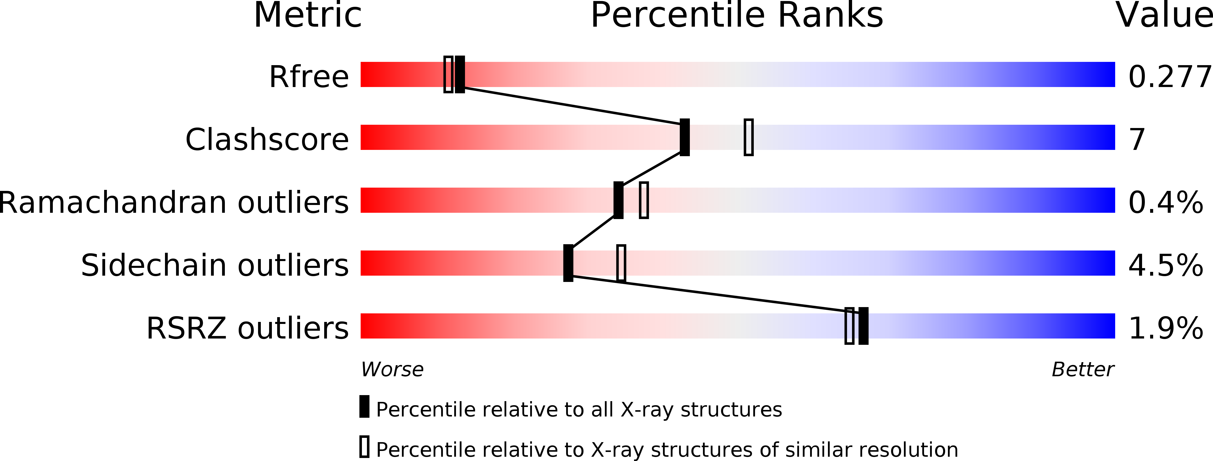
R-Value Free:
0.27
R-Value Work:
0.21
R-Value Observed:
0.21
Space Group:
P 21 21 21