Abstact
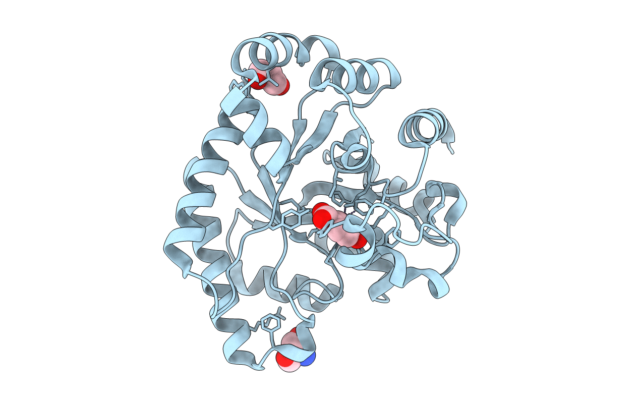
Petrobactin, a virulence-associated siderophore produced by Bacillus anthracis, chelates ferric iron through the rare 3,4-isomer of dihydroxybenzoic acid (3,4-DHBA). Most catechol siderophores, including bacillibactin and enterobactin, use 2,3-DHBA as a biosynthetic subunit. Significantly, siderocalin, a factor involved in human innate immunity, sequesters ferric siderophores bearing the more typical 2,3-DHBA moiety, thereby impeding uptake of iron by the pathogenic bacterial cell. In contrast, the unusual 3,4-DHBA component of petrobactin renders the siderocalin system incapable of obstructing bacterial iron uptake. Although recent genetic and biochemical studies have revealed selected early steps in petrobactin biosynthesis, the origin of 3,4-DHBA as well as the function of the protein encoded by the final gene in the B. anthracis siderophore biosynthetic (asb) operon, asbF (BA1986), has remained unclear. In this study we demonstrate that 3,4-DHBA is produced through conversion of the common bacterial metabolite 3-dehydroshikimate (3-DHS) by AsbF-a 3-DHS dehydratase. Elucidation of the cocrystal structure of AsbF with 3,4-DHBA, in conjunction with a series of biochemical studies, supports a mechanism in which an enolate intermediate is formed through the action of this 3-DHS dehydratase metalloenzyme. Structural and functional parallels are evident between AsbF and other enzymes within the xylose isomerase TIM-barrel family. Overall, these data indicate that microbial species shown to possess homologs of AsbF may, like B. anthracis, also rely on production of the unique 3,4-DHBA metabolite to achieve full viability in the environment or virulence within the host.


