3DWM image
Deposition Date 2008-07-22
Release Date 2008-09-23
Last Version Date 2023-08-30
Entry Detail
PDB ID:
3DWM
Keywords:
Title:
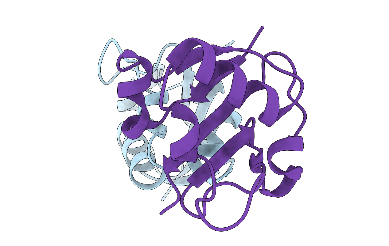
Crystal structure of Mycobacterium tuberculosis CysO, an antigen
Biological Source:
Source Organism:
Host Organism:
Method Details:
Experimental Method:
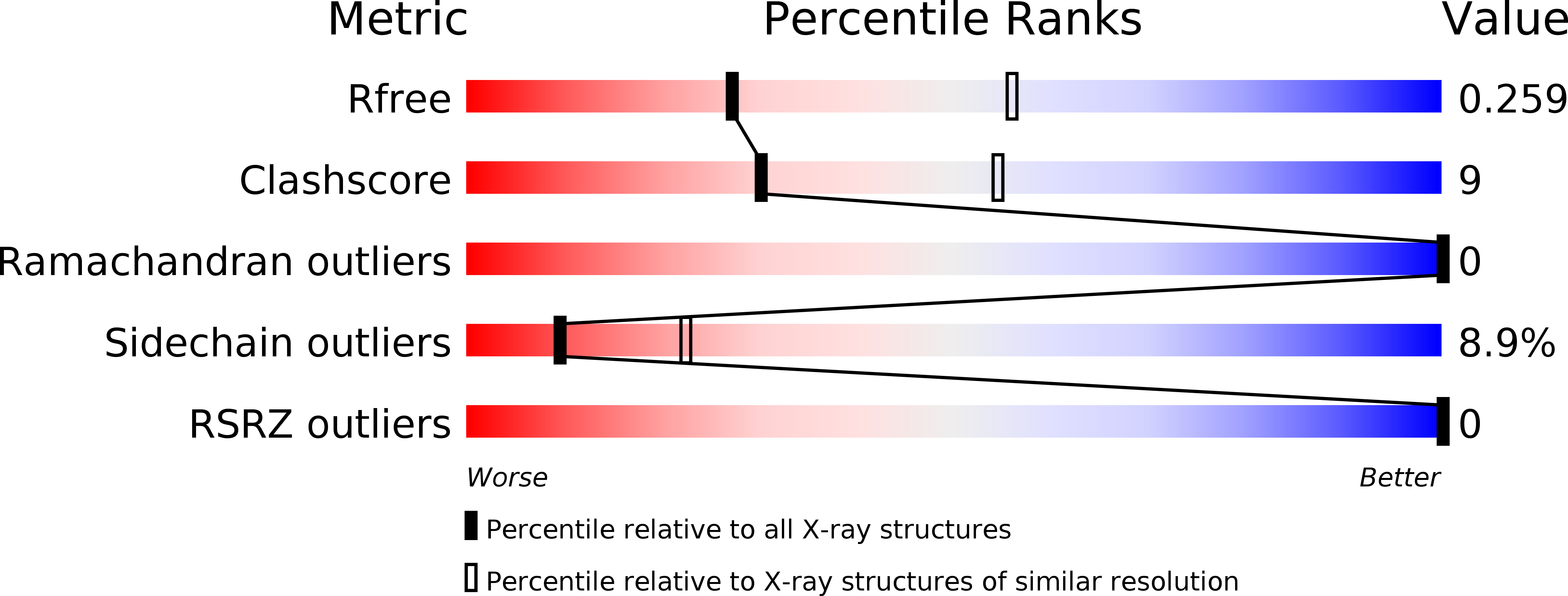
Resolution:
2.69 Å
R-Value Free:
0.25
R-Value Work:
0.19
R-Value Observed:
0.19
Space Group:
C 1 2 1
Macromolecular Entities
Polymer Type:polypeptide(L)
Molecule:9.5 kDa culture filtrate antigen cfp10A
Chain IDs:A, B
Chain Length:93
Number of Molecules:2
Biological Source:Mycobacterium tuberculosis
Primary Citation
Crystal structure of a sulfur carrier protein complex found in the cysteine biosynthetic pathway of Mycobacterium tuberculosis.
Biochemistry 47 10354 10364 (2008)
PMID: 18771296 DOI: 10.1021/bi800915j

Abstact

The structure of the protein complex CysM-CysO from a new cysteine biosynthetic pathway found in the H37Rv strain of Mycobacterium tuberculosis has been determined at 1.53 A resolution. CysM (Rv1336) is a PLP-containing beta-replacement enzyme and CysO (Rv1335) is a sulfur carrier protein with a ubiquitin-like fold. CysM catalyzes the replacement of the acetyl group of O-acetylserine by CysO thiocarboxylate to generate a protein-bound cysteine that is released in a subsequent proteolysis reaction. The protein complex in the crystal structure is asymmetric with one CysO protomer binding to one end of a CysM dimer. Additionally, the structures of CysM and CysO were determined individually at 2.8 and 2.7 A resolution, respectively. Sequence alignments with homologues and structural comparisons with CysK, a cysteine synthase that does not utilize a sulfur carrier protein, revealed high conservation of active site residues; however, residues in CysM responsible for CysO binding are not conserved. Comparison of the CysM-CysO binding interface with other sulfur carrier protein complexes revealed a similarity in secondary structural elements that contribute to complex formation in the ThiF-ThiS and MoeB-MoaD systems, despite major differences in overall folds. Comparison of CysM with and without bound CysO revealed conformational changes associated with CysO binding.

Legend

Protein

Chemical

Disease

Primary Citation of related structures