Deposition Date
2008-07-21
Release Date
2008-07-29
Last Version Date
2024-10-30
Entry Detail
PDB ID:
3DW0
Keywords:
Title:
Crystal structure of the class A carbapenemase KPC-2 at 1.6 angstrom resolution
Biological Source:
Source Organism(s):
Escherichia coli (Taxon ID: 562)
Expression System(s):
Method Details:
Experimental Method:
Resolution:
1.60 Å
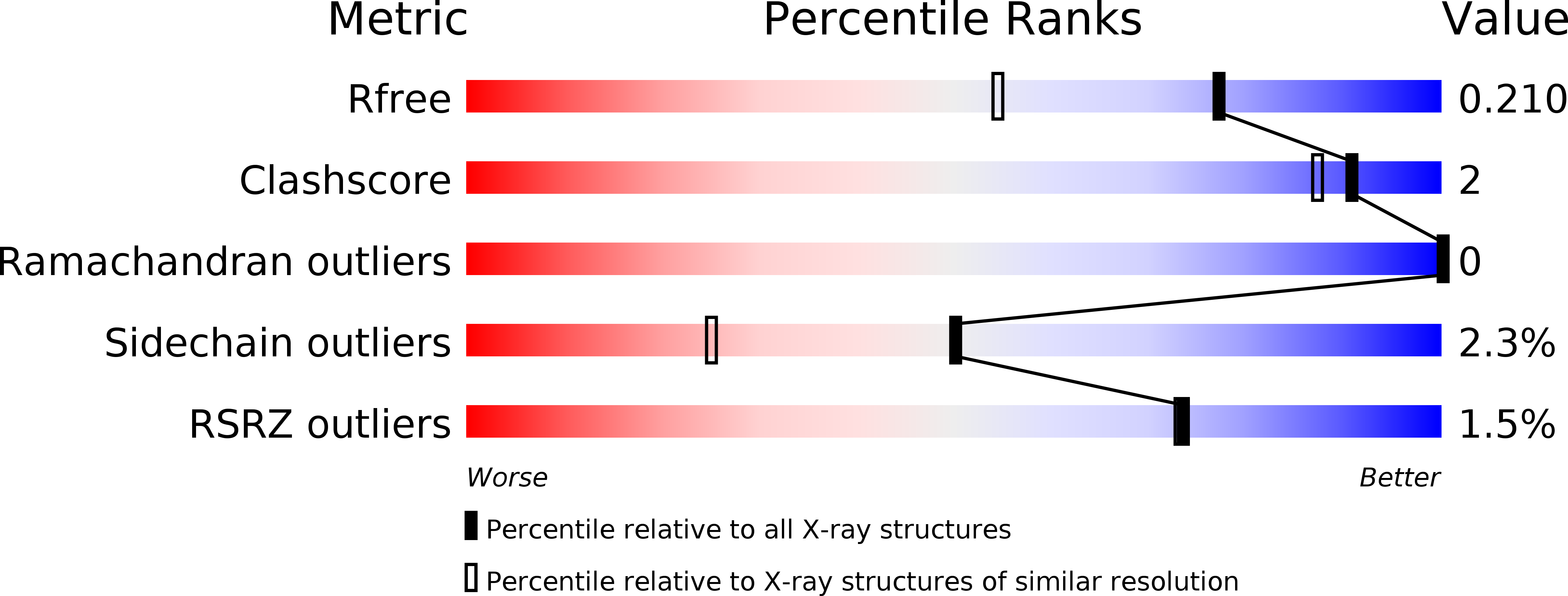
R-Value Free:
0.21
R-Value Work:
0.18
R-Value Observed:
0.18
Space Group:
P 1 21 1