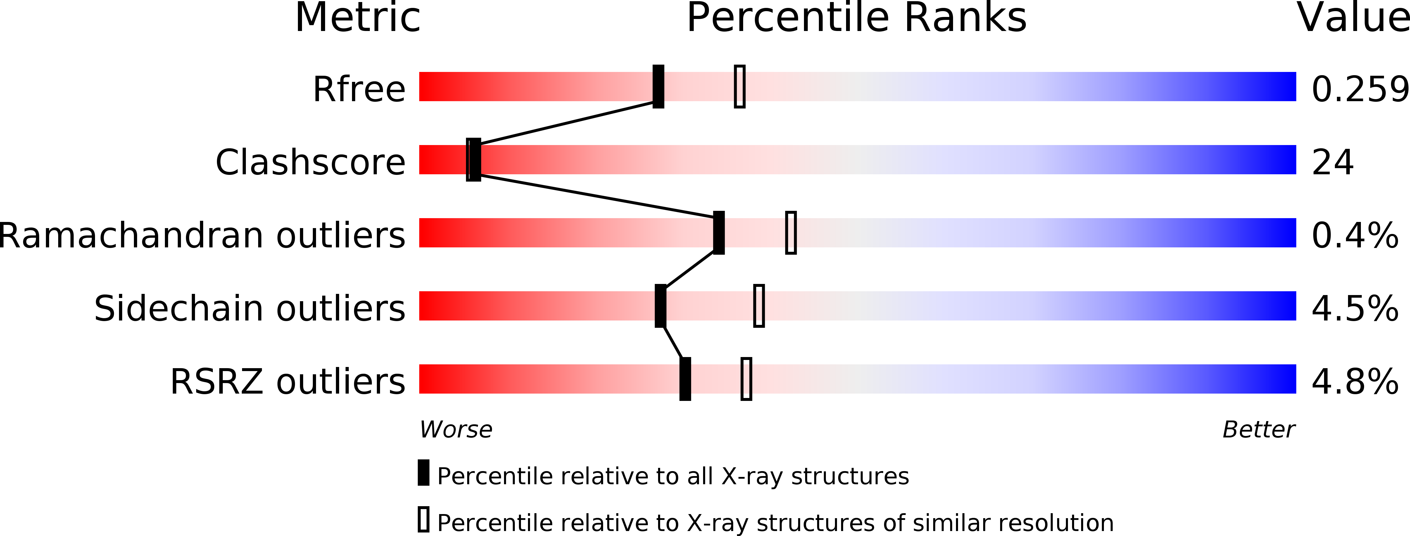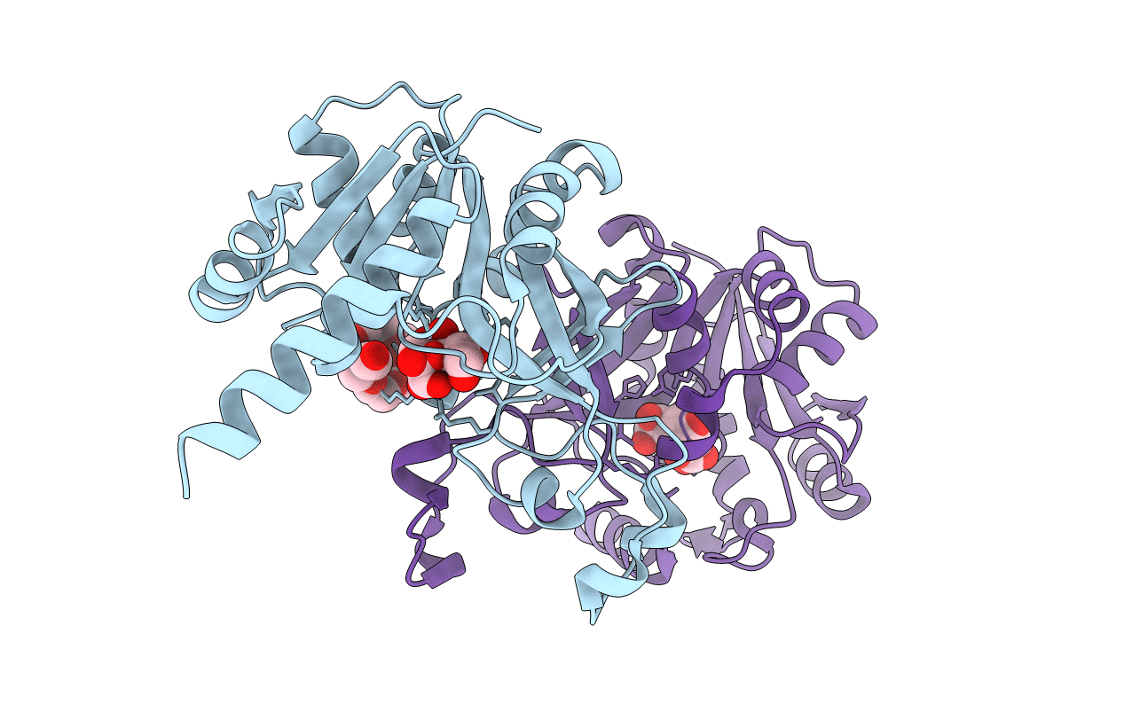
3DUV image
Deposition Date 2008-07-18
Release Date 2008-12-09
Last Version Date 2024-10-09
Entry Detail
PDB ID:
3DUV
Keywords:
Title:
Crystal structure of 3-deoxy-manno-octulosonate cytidylyltransferase from Haemophilus influenzae complexed with the substrate 3-deoxy-manno-octulosonate in the-configuration
Biological Source:
Source Organism:
Host Organism:
Method Details:
Experimental Method:
Resolution:
2.30 Å
R-Value Free:
0.23
R-Value Work:
0.17
R-Value Observed:
0.17
Space Group:
P 21 21 21
Macromolecular Entities
Polymer Type:polypeptide(L)
Molecule:3-deoxy-manno-octulosonate cytidylyltransferase
Gene (Uniprot):kdsB
Chain IDs:A, B
Chain Length:262
Number of Molecules:2
Biological Source:Haemophilus influenzae
Primary Citation
Structure of 3-deoxy-manno-octulosonate cytidylyltransferase from Haemophilus influenzae complexed with the substrate 3-deoxy-manno-octulosonate in the beta-configuration.
Acta Crystallogr.,Sect.D 64 1292 1294 (2008)
PMID: 19018107 DOI: 10.1107/S0907444908036342

Abstact

The enzyme 3-deoxy-manno-octulosonate cytidylyltransferase (CMP-KDO synthetase; CKS) catalyzes the activation of 3-deoxy-D-manno-octulosonate (or 2-keto-3-deoxy-manno-octonic acid; KDO) by forming CMP-KDO. CKS is unique to Gram-negative bacteria and is an attractive target for the development of antibacterial agents. The crystal structure of CKS from Haemophilus influenzae in complex with the substrate KDO has been determined at 2.30 A resolution by combining single-wavelength anomalous diffraction and molecular-replacement methods. The two monomers in the asymmetric unit differ in the conformation of their C-terminal alpha-helix (Ala230-Asn254). The KDO bound to the active site exists as the beta-pyranose form in the (5)C(2) chair conformation. The structure of CKS from H. influenzae in complex with KDO will be useful in structure-based inhibitor design.

Legend

Protein

Chemical

Disease

Primary Citation of related structures