
Deposition Date
2008-07-13
Release Date
2009-03-10
Last Version Date
2024-03-20
Entry Detail
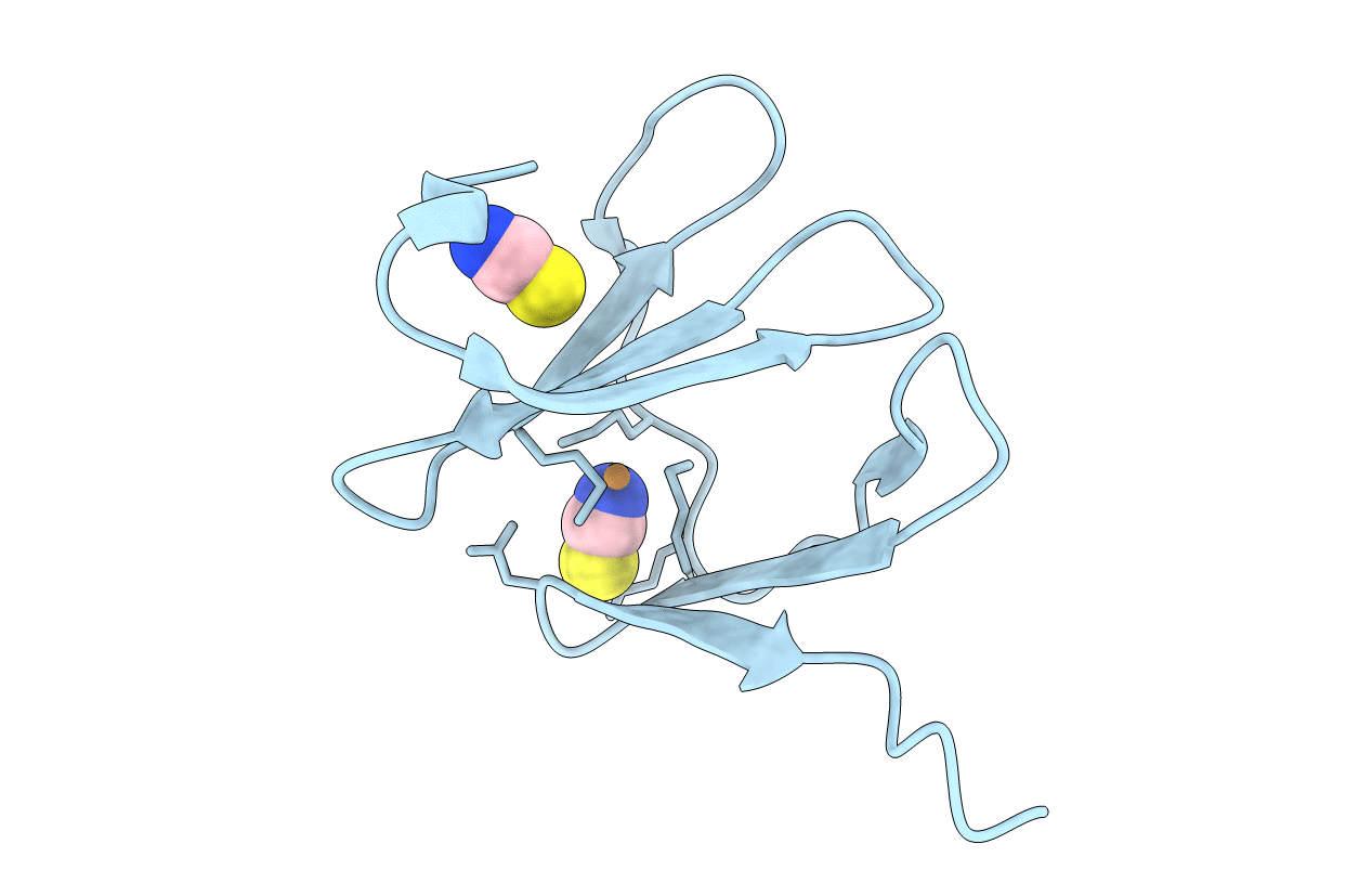
PDB ID:
3DSO
Keywords:
Title:
Crystal structure of Cu(I) bound copper resistance protein CopK
Biological Source:
Source Organism(s):
Ralstonia metallidurans (Taxon ID: 266264)
Expression System(s):
Method Details:
Experimental Method:
Resolution:
1.55 Å
R-Value Free:
0.21
R-Value Work:
0.20
R-Value Observed:
0.20
Space Group:
P 21 21 2
