3DSL image
Deposition Date 2008-07-13
Release Date 2008-10-21
Last Version Date 2024-11-20
Entry Detail
PDB ID:
3DSL
Keywords:
Title:
The Three-dimensional Structure of Bothropasin, the Main Hemorrhagic Factor from Bothrops jararaca venom.
Biological Source:
Source Organism:
Method Details:
Experimental Method:
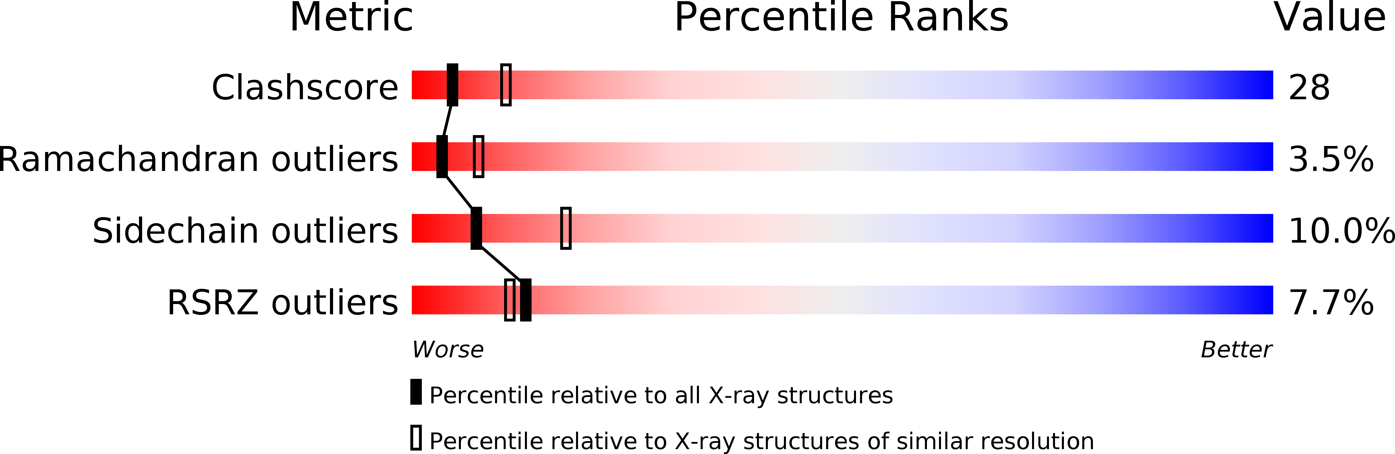
Resolution:
2.70 Å
R-Value Free:
0.29
R-Value Work:
0.21
R-Value Observed:
0.21
Space Group:
P 21 21 21
Macromolecular Entities
Structures with similar UniProt ID
Protein Blast
Polymer Type:polypeptide(L)
Molecule:Zinc metalloproteinase-disintegrin bothropasin
Chain IDs:A, B
Chain Length:419
Number of Molecules:2
Biological Source:Bothrops jararaca
Modified Residue
Compound ID Chain ID Parent Comp ID Details 2D Image
ASN A ASN GLYCOSYLATION SITE
Primary Citation
The three-dimensional structure of bothropasin, the main hemorrhagic factor from Bothrops jararaca venom: Insights for a new classification of snake venom metalloprotease subgroups.
Toxicon 52 807 816 (2008)
PMID: 18831982 DOI: 10.1016/j.toxicon.2008.08.021

Abstact

Bothropasin is a 48kDa hemorrhagic PIII snake venom metalloprotease (SVMP) isolated from Bothrops jararaca, containing disintegrin/cysteine-rich adhesive domains. Here we present the crystal structure of bothropasin complexed with the inhibitor POL647. The catalytic domain consists of a scaffold of two subdomains organized similarly to those described for other SVMPs, including the zinc and calcium-binding sites. The free cysteine residue Cys189 is located within a hydrophobic core and it is not available for disulfide bonding or other interactions. There is no identifiable secondary structure for the disintegrin domain, but instead it is composed mostly of loops stabilized by seven disulfide bonds and by two calcium ions. The ECD region is in a loop and is structurally related to the RGD region of RGD disintegrins, which are derived from PII SVMPs. The ECD motif is stabilized by the Cys277-Cys310 disulfide bond (between the disintegrin and cysteine-rich domains) and by one calcium ion. The side chain of Glu276 of the ECD motif is exposed to solvent and free to make interactions. In bothropasin, the HVR (hyper-variable region) described for other PIII SVMPs in the cysteine-rich domain, presents a well-conserved sequence with respect to several other PIII members from different species. We propose that this subset be referred to as PIII-HCR (highly conserved region) SVMPs. The differences in the disintegrin-like, cysteine-rich or disintegrin-like cysteine-rich domains may be involved in selecting target binding, which in turn could generate substrate diversity or specificity for the catalytic domain.

Legend

Protein

Chemical

Disease

Primary Citation of related structures
Feedback Form
Name
Email
Institute
Feedback