Deposition Date
2008-07-12
Release Date
2008-10-07
Last Version Date
2024-02-21
Entry Detail
PDB ID:
3DSH
Keywords:
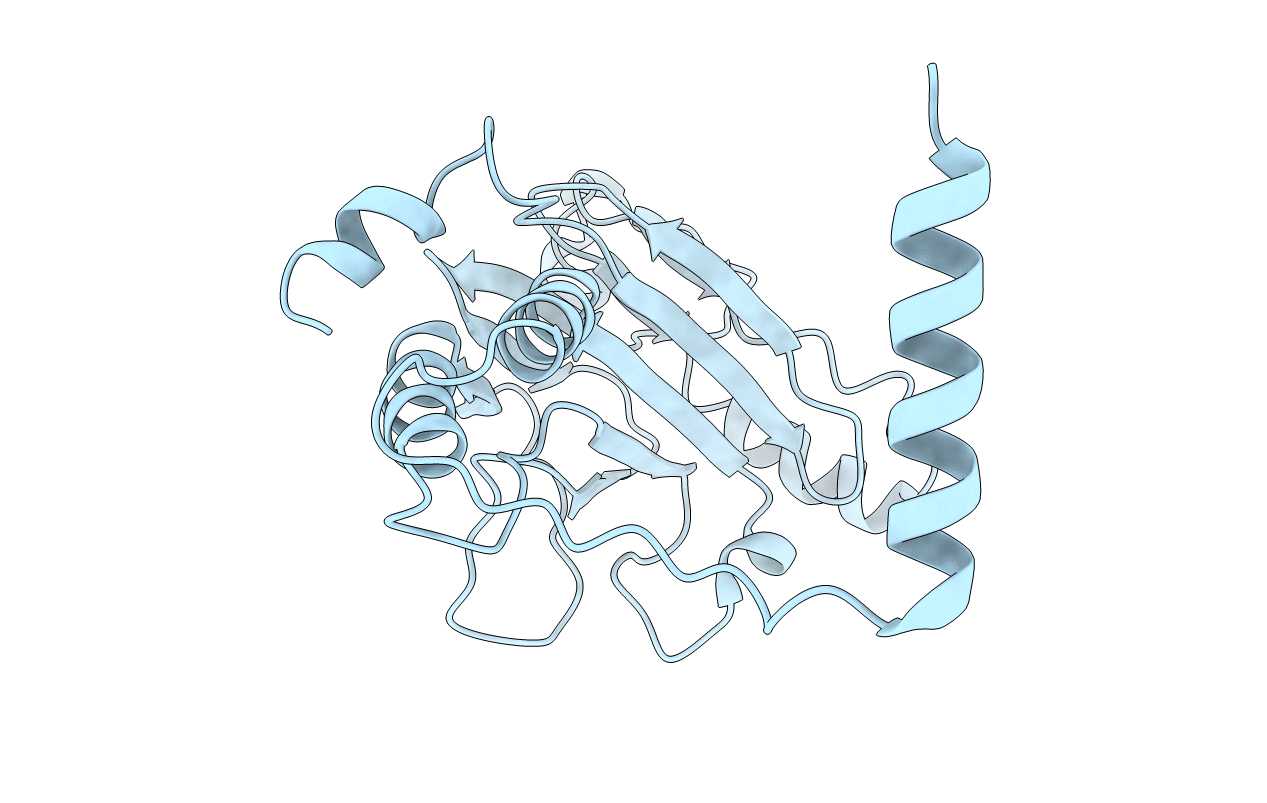
Title:
Crystal structure of dimeric interferon regulatory factor 5 (IRF-5) transactivation domain
Biological Source:
Source Organism(s):
Homo sapiens (Taxon ID: 9606)
Expression System(s):
Method Details:
Experimental Method:
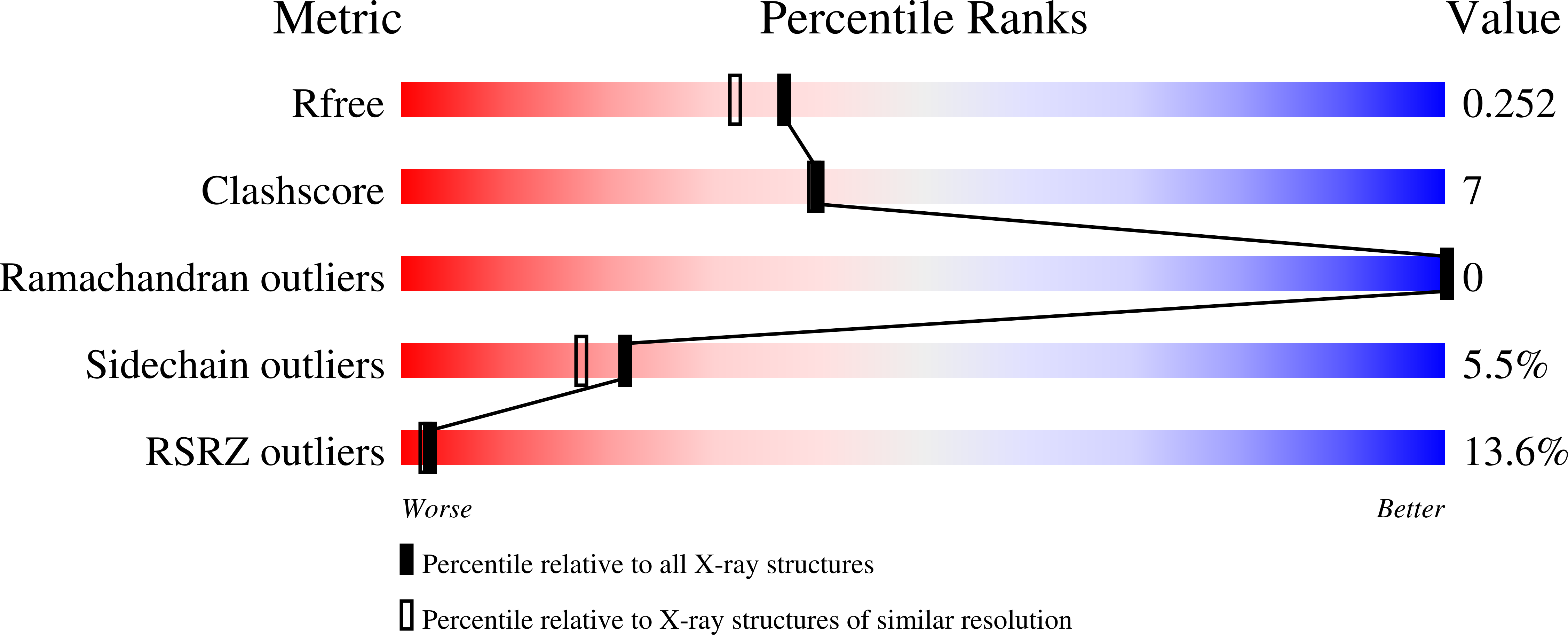
Resolution:
2.00 Å
R-Value Free:
0.24
R-Value Work:
0.20
R-Value Observed:
0.20
Space Group:
P 31 2 1