Deposition Date
2008-07-07
Release Date
2008-09-09
Last Version Date
2024-05-29
Entry Detail
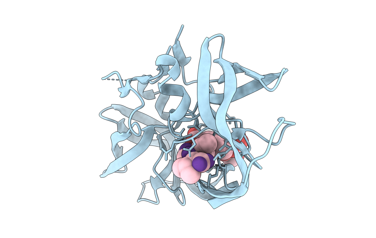
Biological Source:
Source Organism(s):
Expression System(s):
Method Details:
Experimental Method:
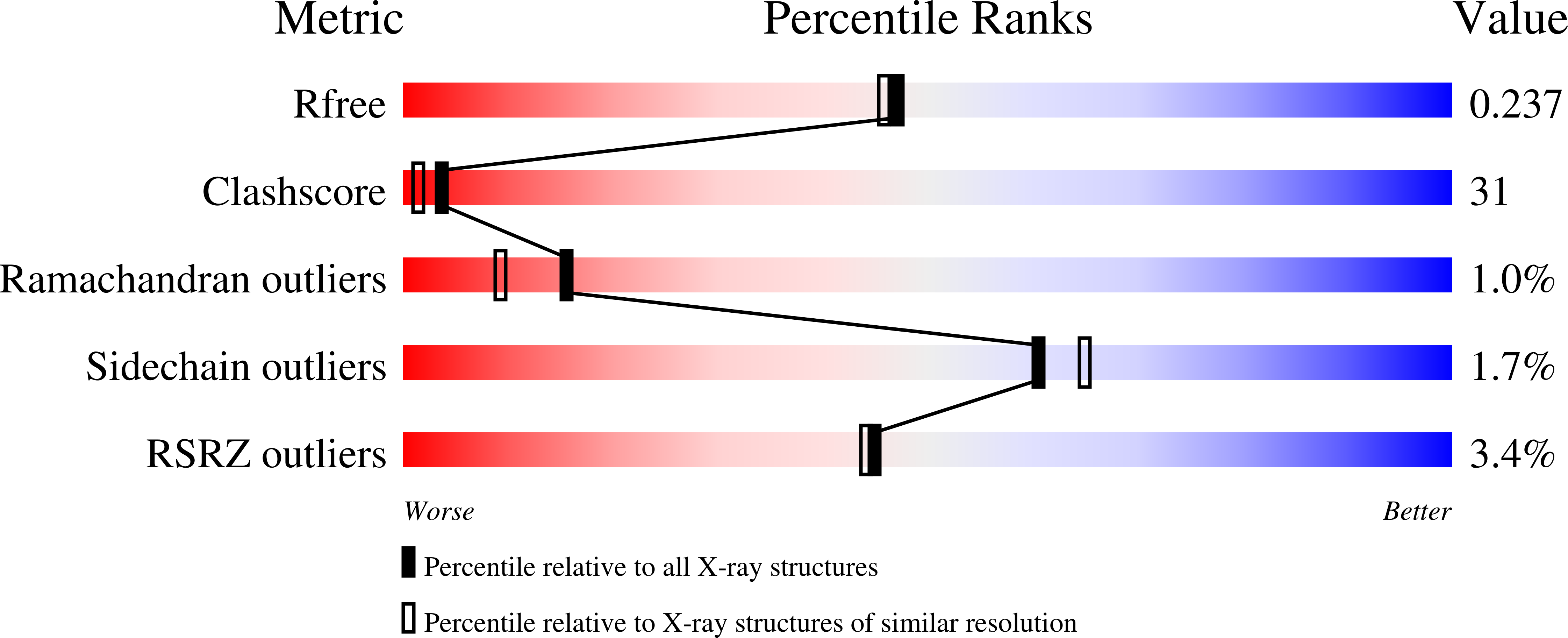
Resolution:
2.00 Å
R-Value Free:
0.23
R-Value Work:
0.19
R-Value Observed:
0.19
Space Group:
P 61