3DOO image
Deposition Date 2008-07-05
Release Date 2009-05-05
Last Version Date 2023-11-01
Entry Detail
PDB ID:
3DOO
Keywords:
Title:
Crystal structure of shikimate dehydrogenase from Staphylococcus epidermidis complexed with shikimate
Biological Source:
Source Organism:
Host Organism:
Method Details:
Experimental Method:
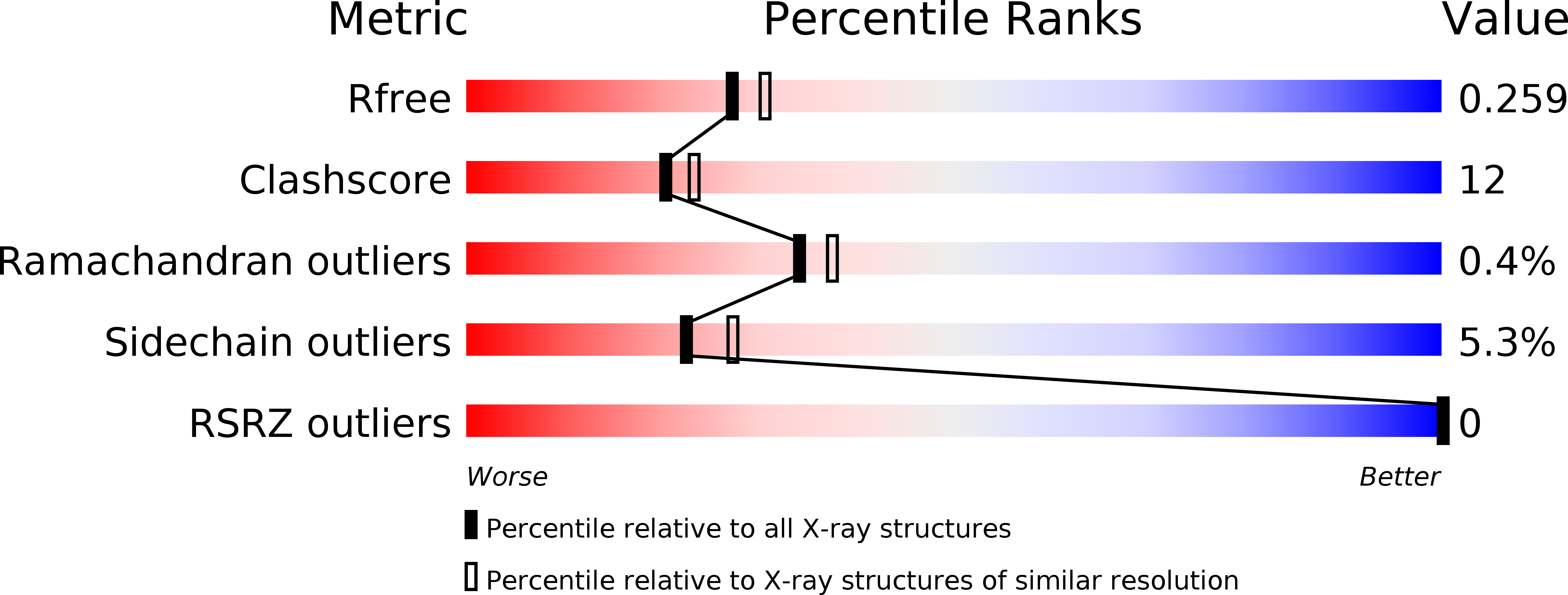
Resolution:
2.20 Å
R-Value Free:
0.26
R-Value Work:
0.18
R-Value Observed:
0.19
Space Group:
P 1 21 1
Macromolecular Entities
Polymer Type:polypeptide(L)
Molecule:Shikimate dehydrogenase
Gene (Uniprot):aroE
Chain IDs:A
Chain Length:277
Number of Molecules:1
Biological Source:Staphylococcus epidermidis
Ligand Molecules
Primary Citation
X-ray crystallographic and enzymatic analyses of shikimate dehydrogenase from Staphylococcus epidermidis
Febs J. 276 1125 1139 (2009)
PMID: 19215302 DOI: 10.1111/j.1742-4658.2008.06856.x

Abstact

Shikimate dehydrogenase (SDH) catalyzes the NADPH-dependent reduction of 3-dehydroshikimate to shikimate in the shikimate pathway. In this study, we determined the kinetic properties and crystal structures of Staphylococcus epidermidis SDH (SeSDH) both in its ligand-free form and in complex with shikimate. SeSDH has a k(cat) of 22.8 s(-1) and a K(m) of 73 mum towards shikimate, and a K(m) of 100 microM towards NADP. The overall folding of SeSDH comprises the N-terminal alpha/beta domain for substrate binding and the C-terminal Rossmann fold for NADP binding. The active site is within a large groove between the two domains. Residue Tyr211, normally regarded as important for substrate binding, does not interact with shikimate in the binary SeSDH-shikimate complex structure. However, the Y211F mutation leads to a significant decrease in k(cat) and a minor increase in the K(m) for shikimate. The results indicate that the main function of Tyr211 may be to stabilize the catalytic intermediate during catalysis. The NADP-binding domain of SeSDH is less conserved. The usually long helix specifically recognizing the adenine ribose phosphate is substituted with a short 3(10) helix in the NADP-binding domain. Moreover, the interdomain angle of SeSDH is the widest among all known SDH structures, indicating an inactive 'open' state of the SeSDH structure. Thus, a 'closing' process might occur upon NADP binding to bring the cofactor close to the substrate for catalysis.

Legend

Protein

Chemical

Disease

Primary Citation of related structures