3DLA image
Deposition Date 2008-06-26
Release Date 2009-03-10
Last Version Date 2025-03-26
Entry Detail
PDB ID:
3DLA
Keywords:
Title:
X-ray crystal structure of glutamine-dependent NAD+ synthetase from Mycobacterium tuberculosis bound to NaAD+ and DON
Biological Source:
Source Organism:
Host Organism:
Method Details:
Experimental Method:
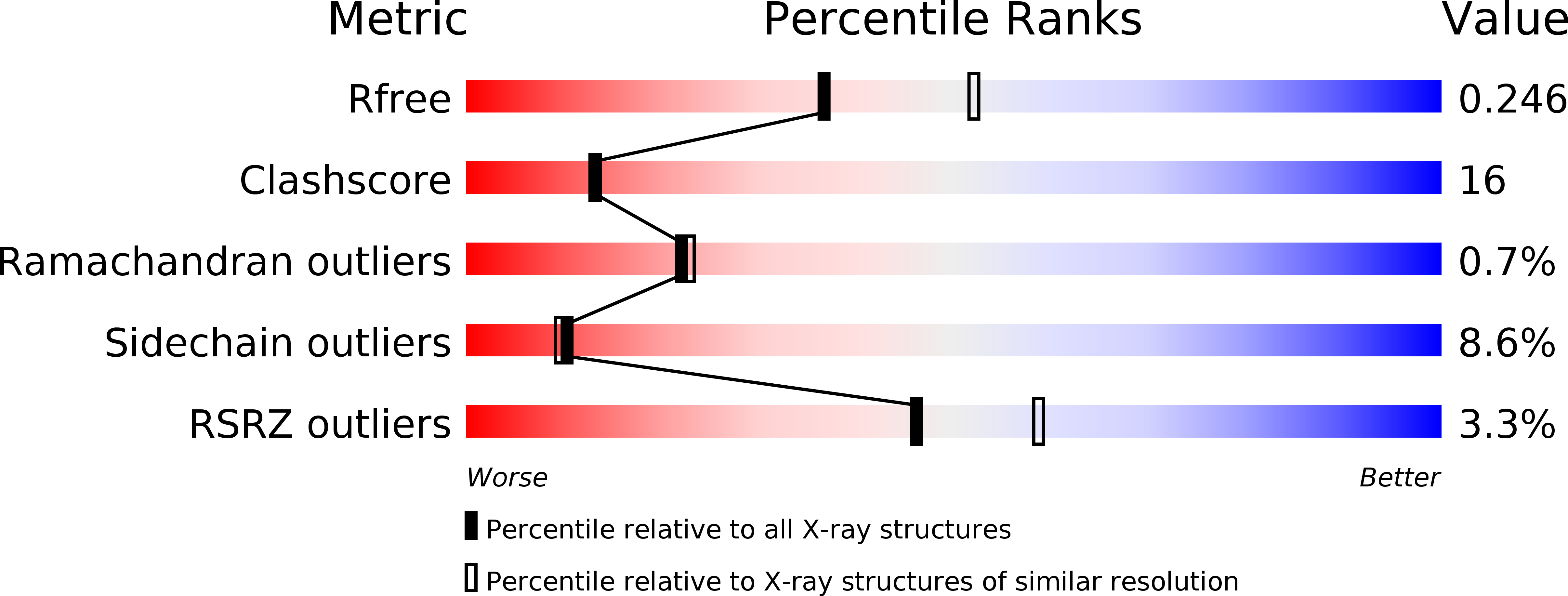
Resolution:
2.35 Å
R-Value Free:
0.24
R-Value Work:
0.18
R-Value Observed:
0.18
Space Group:
P 41 21 2
Macromolecular Entities
Structures with similar UniProt ID
Protein Blast
Polymer Type:polypeptide(L)
Molecule:Glutamine-dependent NAD(+) synthetase
Chain IDs:A, B, C, D
Chain Length:680
Number of Molecules:4
Biological Source:Mycobacterium tuberculosis
Primary Citation
Regulation of active site coupling in glutamine-dependent NAD(+) synthetase.
Nat.Struct.Mol.Biol. 16 421 429 (2009)
PMID: 19270703 DOI: 10.1038/nsmb.1567

Abstact

NAD(+) is an essential metabolite both as a cofactor in energy metabolism and redox homeostasis and as a regulator of cellular processes. In contrast to humans, Mycobacterium tuberculosis NAD(+) biosynthesis is absolutely dependent on the activity of a multifunctional glutamine-dependent NAD(+) synthetase, which catalyzes the ATP-dependent formation of NAD(+) at the synthetase domain using ammonia derived from L-glutamine in the glutaminase domain. Here we report the kinetics and structural characterization of M. tuberculosis NAD(+) synthetase. The kinetics data strongly suggest tightly coupled regulation of the catalytic activities. The structure, the first of a glutamine-dependent NAD(+) synthetase, reveals a homooctameric subunit organization suggesting a tight dependence of catalysis on the quaternary structure, a 40-A intersubunit ammonia tunnel and structural elements that may be involved in the transfer of information between catalytic sites.

Legend

Protein

Chemical

Disease

Primary Citation of related structures
Feedback Form
Name
Email
Institute
Feedback