3DI1 image
Deposition Date 2008-06-19
Release Date 2008-08-12
Last Version Date 2024-10-30
Entry Detail
PDB ID:
3DI1
Keywords:
Title:
Crystal structure of the Staphylococcus aureus Dihydrodipicolinate synthase-pyruvate complex
Biological Source:
Source Organism:
Host Organism:
Method Details:
Experimental Method:
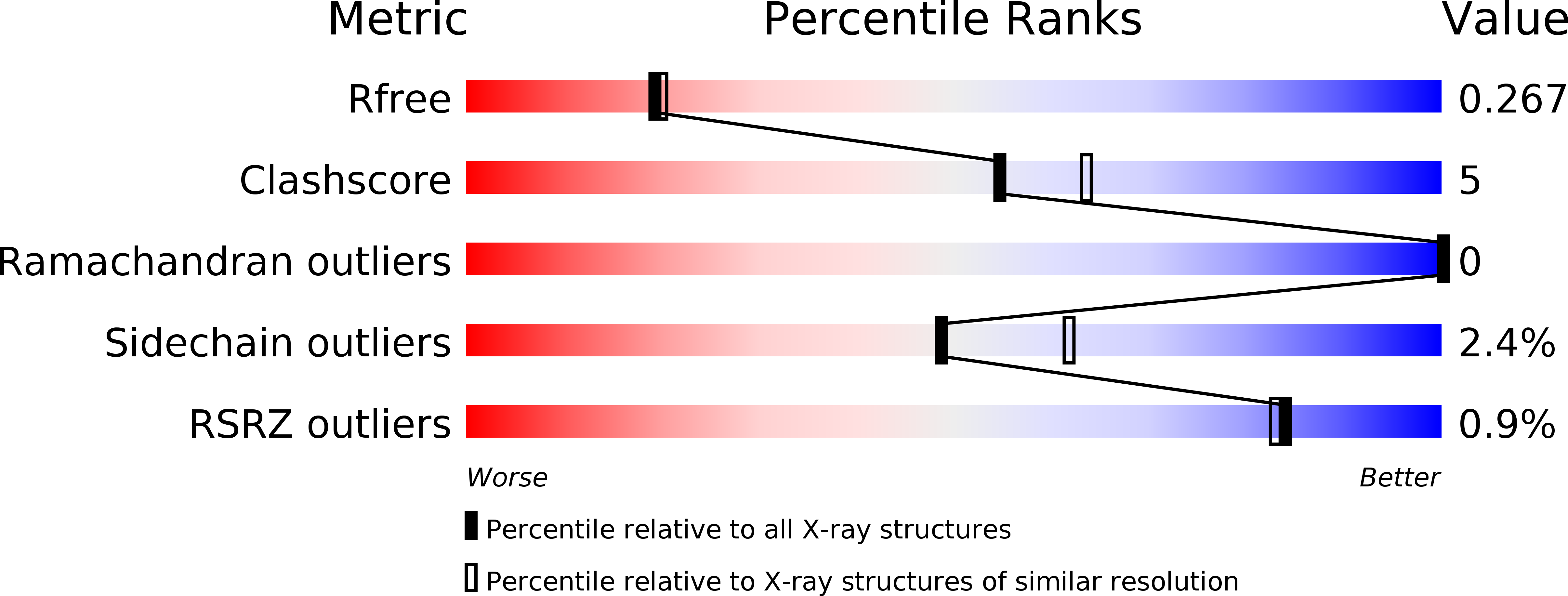
Resolution:
2.20 Å
R-Value Free:
0.23
R-Value Work:
0.18
R-Value Observed:
0.19
Space Group:
P 64 2 2
Macromolecular Entities
Structures with similar UniProt ID
Protein Blast
Polymer Type:polypeptide(L)
Molecule:Dihydrodipicolinate synthase
Gene (Uniprot):dapA
Chain IDs:A, B
Chain Length:295
Number of Molecules:2
Biological Source:Staphylococcus aureus subsp. aureus
Ligand Molecules
Primary Citation
Structural and functional characterization of Staphylococcus aureus dihydrodipicolinate synthase
Febs Lett. 582 2923 2930 (2008)
PMID: 18671976 DOI: 10.1016/j.febslet.2008.07.035

Abstact

Lysine biosynthesis is crucial for cell-wall formation in bacteria. Enzymes involved in lysine biosynthesis are thus potential targets for anti-microbial therapeutics. Dihydrodipicolinate synthase (DHDPS) catalyzes the first step of this pathway. Unlike its homologues, Staphylococcus aureus DHDPS is a dimer both in solution and in the crystal and is not feedback inhibited by lysine. The crystal structure of S. aureus DHDPS in the free and substrate bound forms provides a structural rationale for its catalytic mechanism. The structure also reveals unique conformational features of the S. aureus enzyme that could be crucial for the design of specific non-competitive inhibitors.

Legend

Protein

Chemical

Disease

Primary Citation of related structures