
Deposition Date
2008-06-10
Release Date
2008-11-25
Last Version Date
2024-03-13
Entry Detail
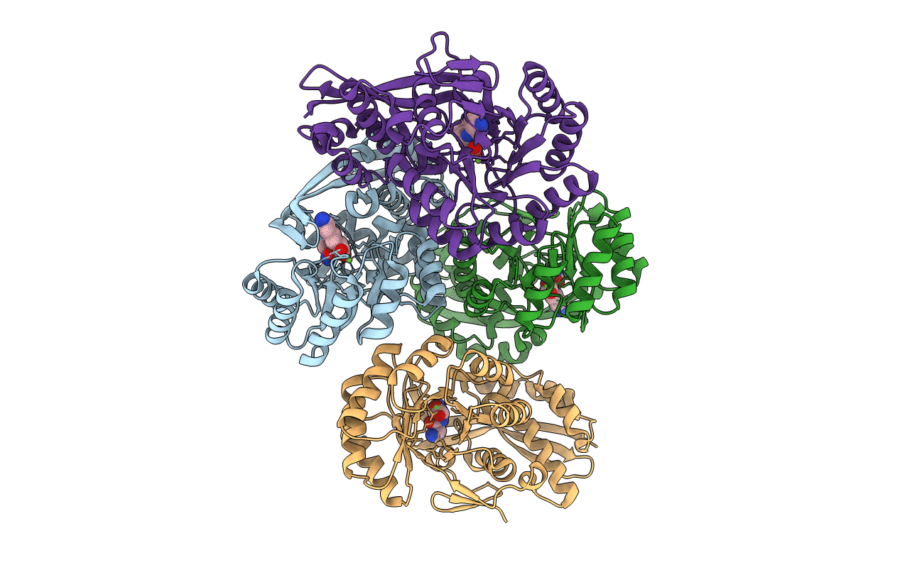
PDB ID:
3DER
Keywords:
Title:
Crystal structure of dipeptide epimerase from Thermotoga maritima complexed with L-Ala-L-Lys dipeptide
Biological Source:
Source Organism(s):
Thermotoga maritima MSB8 (Taxon ID: 243274)
Expression System(s):
Method Details:
Experimental Method:
Resolution:
1.90 Å
R-Value Free:
0.24
R-Value Work:
0.23
R-Value Observed:
0.23
Space Group:
P 61 2 2
