3DDT image
Deposition Date 2008-06-06
Release Date 2008-10-07
Last Version Date 2024-03-20
Entry Detail
PDB ID:
3DDT
Keywords:
Title:
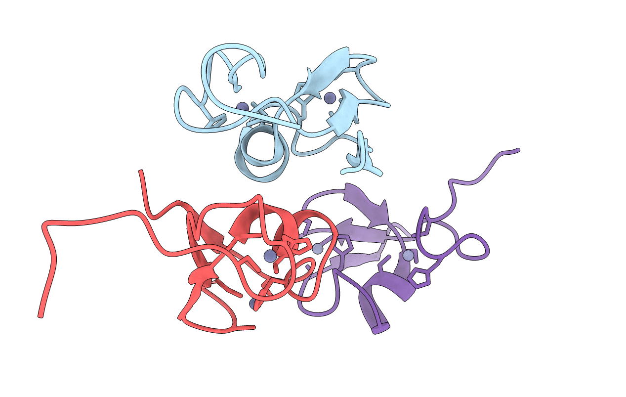
Crystal structure of the B2 box from MuRF1 in dimeric state
Biological Source:
Source Organism:
Homo sapiens (Taxon ID: 9606)
Host Organism:
Method Details:
Experimental Method:
Resolution:
1.90 Å
R-Value Free:
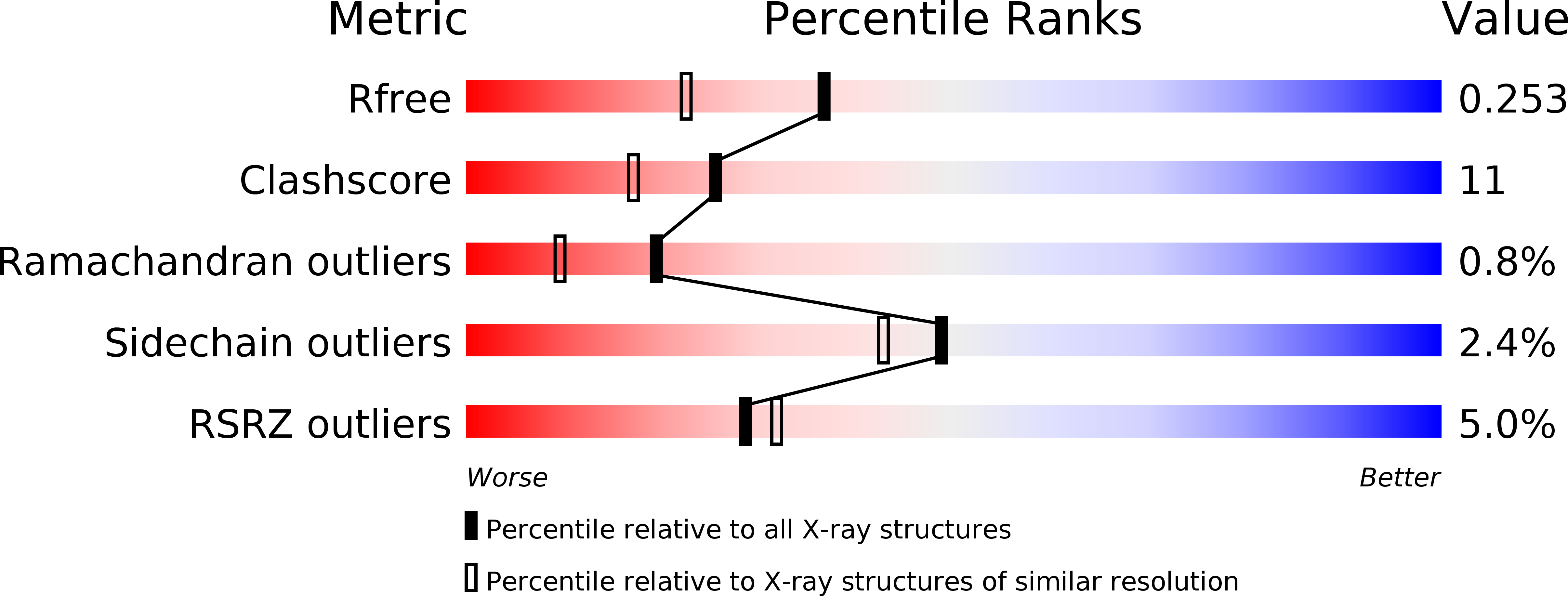
0.25
R-Value Work:
0.19
R-Value Observed:
0.20
Space Group:
P 65 2 2
Macromolecular Entities
Polymer Type:polypeptide(L)
Molecule:E3 ubiquitin-protein ligase TRIM63
Gene (Uniprot):TRIM63
Chain IDs:A, B, C
Chain Length:48
Number of Molecules:3
Biological Source:Homo sapiens
Ligand Molecules
Primary Citation
Structural analysis of B-Box 2 from MuRF1: identification of a novel self-association pattern in a RING-like fold
Biochemistry 47 10722 10730 (2008)
PMID: 18795805 DOI: 10.1021/bi800733z

Abstact

The B-box motif is the defining feature of the TRIM family of proteins, characterized by a RING finger-B-box-coiled coil tripartite fold. We have elucidated the crystal structure of B-box 2 (B2) from MuRF1, a TRIM protein that supports a wide variety of protein interactions in the sarcomere and regulates the trophic state of striated muscle tissue. MuRF1 B2 coordinates two zinc ions through a cross-brace alpha/beta-topology typical of members of the RING finger superfamily. However, it self-associates into dimers with high affinity. The dimerization pattern is mediated by the helical component of this fold and is unique among RING-like folds. This B2 reveals a long shallow groove that encircles the C-terminal metal binding site ZnII and appears as the defining protein-protein interaction feature of this domain. A cluster of conserved hydrophobic residues in this groove and, in particular, a highly conserved aromatic residue (Y133 in MuRF1 B2) is likely to be central to this role. We expect these findings to aid the future exploration of the cellular function and therapeutic potential of MuRF1.

Legend

Protein

Chemical

Disease

Primary Citation of related structures