Deposition Date
2008-06-05
Release Date
2008-09-23
Last Version Date
2023-08-30
Entry Detail
PDB ID:
3DDK
Keywords:
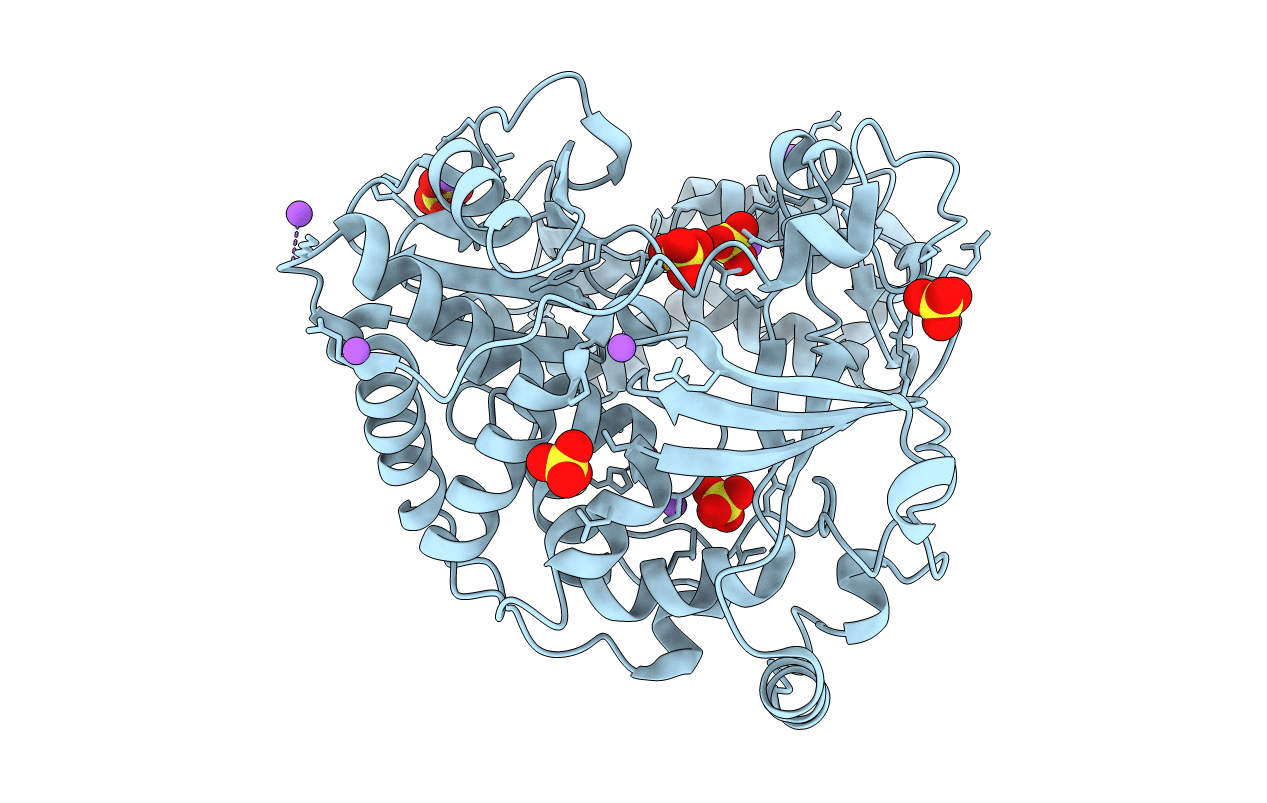
Title:
Coxsackievirus B3 3Dpol RNA Dependent RNA Polymerase
Biological Source:
Source Organism(s):
Human coxsackievirus (Taxon ID: 12072)
Expression System(s):
Method Details:
Experimental Method:
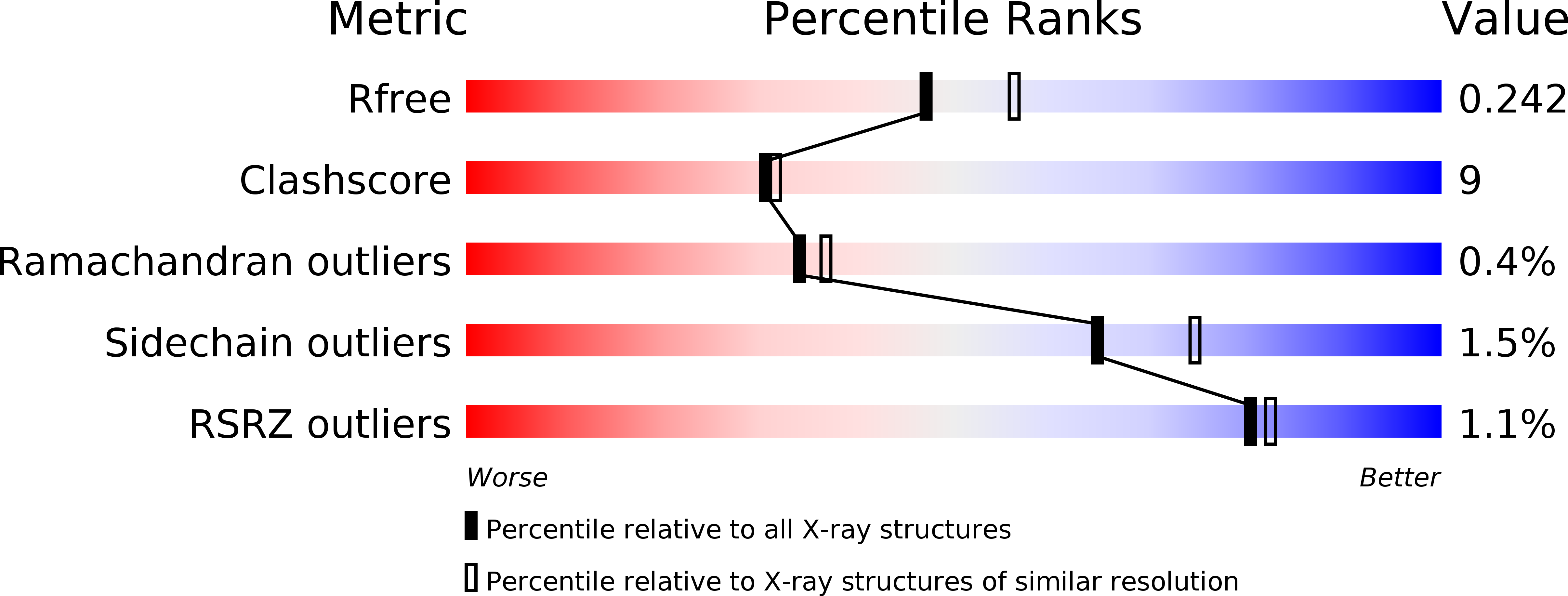
Resolution:
2.25 Å
R-Value Free:
0.23
R-Value Work:
0.20
R-Value Observed:
0.20
Space Group:
P 43 21 2