Deposition Date
2008-06-03
Release Date
2009-06-09
Last Version Date
2023-11-01
Entry Detail
PDB ID:
3DC5
Keywords:
Title:
Crystal Structure of a manganese superoxide dismutases from Caenorhabditis elegans
Biological Source:
Source Organism(s):
Caenorhabditis elegans (Taxon ID: 6239)
Expression System(s):
Method Details:
Experimental Method:
Resolution:
1.70 Å
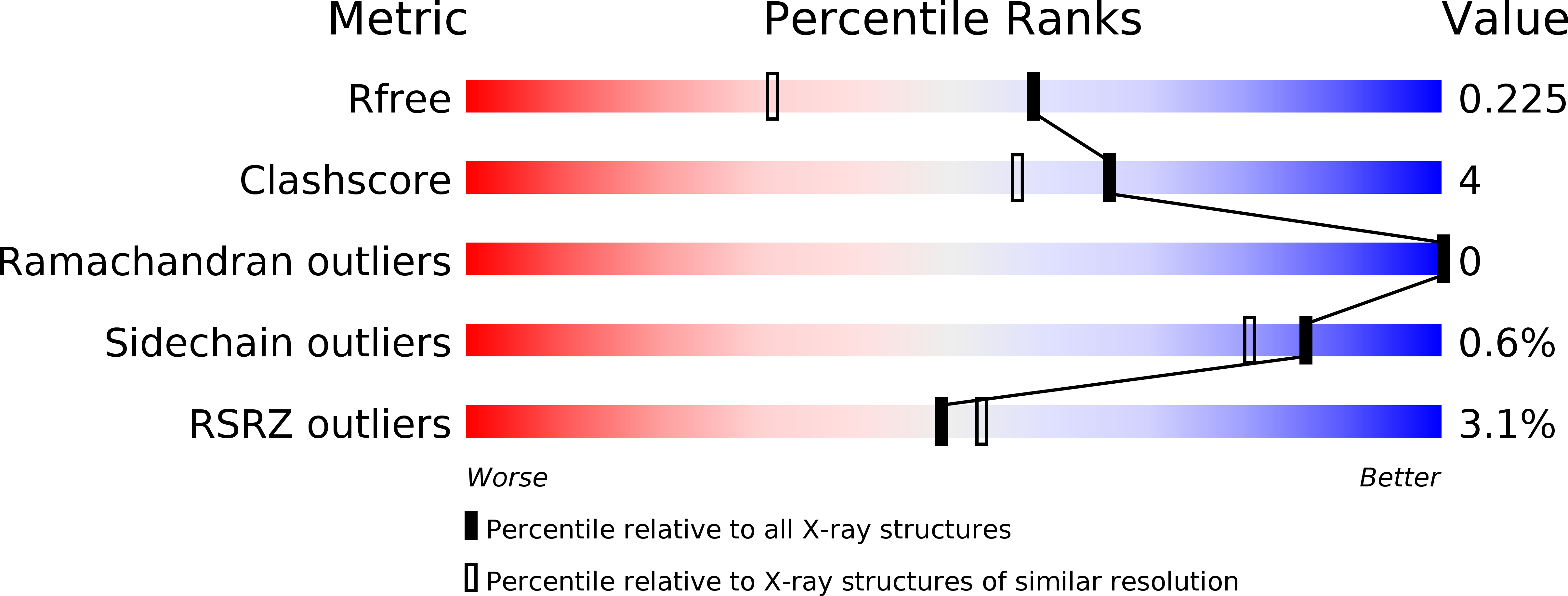
R-Value Free:
0.22
R-Value Work:
0.18
R-Value Observed:
0.19
Space Group:
P 41 21 2