3DAD image
Deposition Date 2008-05-29
Release Date 2008-09-16
Last Version Date 2024-02-21
Entry Detail
PDB ID:
3DAD
Title:
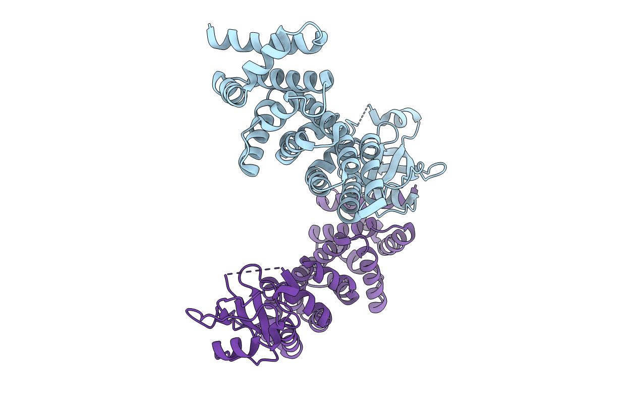
Crystal structure of the N-terminal regulatory domains of the formin FHOD1
Biological Source:
Source Organism:
Homo sapiens (Taxon ID: 9606)
Host Organism:
Method Details:
Experimental Method:
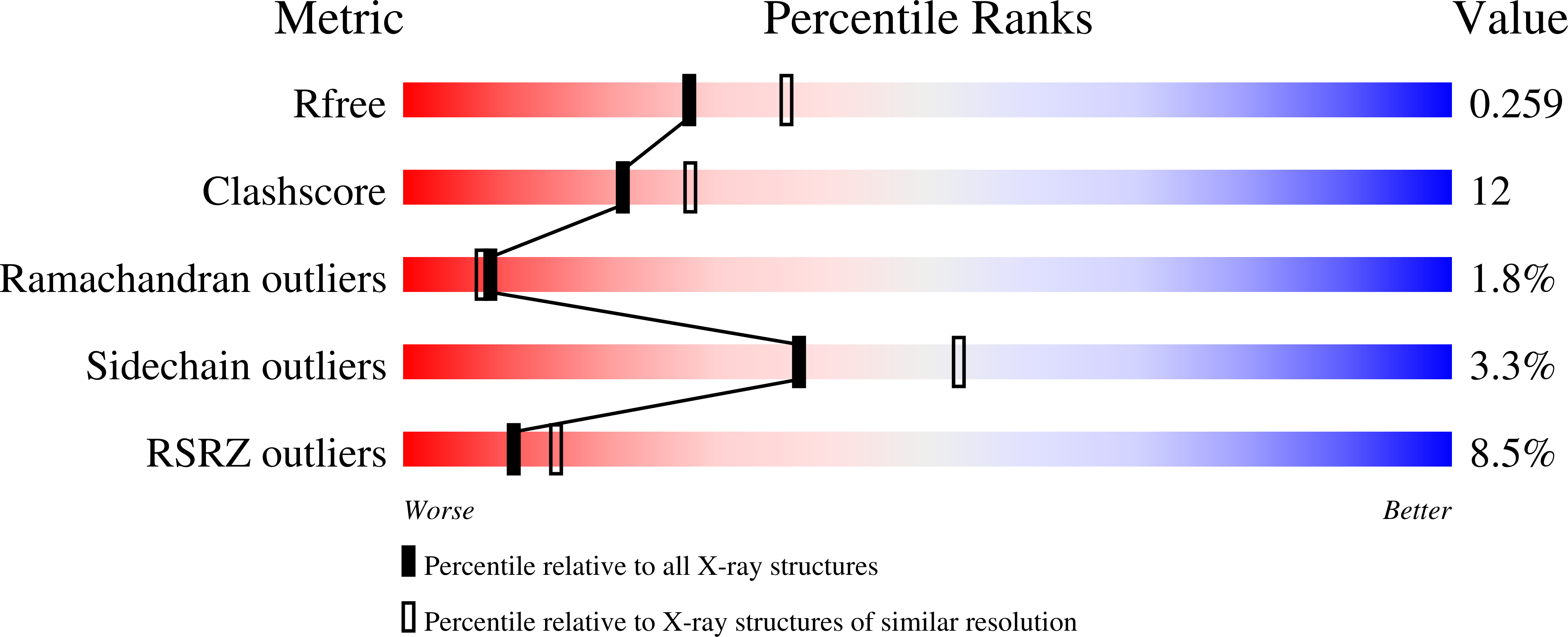
Resolution:
2.30 Å
R-Value Free:
0.25
R-Value Work:
0.21
Space Group:
P 1
Macromolecular Entities
Polymer Type:polypeptide(L)
Molecule:FH1/FH2 domain-containing protein 1
Gene (Uniprot):FHOD1
Mutations:C31S, C71S
Chain IDs:A, B
Chain Length:339
Number of Molecules:2
Biological Source:Homo sapiens
Primary Citation
The Human Formin FHOD1 Contains a Bipartite Structure of FH3 and GTPase-Binding Domains Required for Activation.
Structure 16 1313 1323 (2008)
PMID: 18786395 DOI: 10.1016/j.str.2008.06.008

Abstact

Formins induce the nucleation and polymerization of unbranched actin filaments. They share three homology domains required for profilin binding, actin polymerization, and regulation. Diaphanous-related formins (DRFs) are activated by GTPases of the Rho/Rac family, whose interaction with the N-terminal formin domain is thought to displace a C-terminal Diaphanous-autoregulatory domain (DAD). We have determined the structure of the N-terminal domains of FHOD1 consisting of a GTPase-binding domain (GBD) and the DAD-recognition domain FH3. In contrast to the formin mDia1, the FHOD1-GBD reveals a ubiquitin superfold as found similarly in c-Raf1 or PI3 kinase. This GBD is recruited by Rac and Ras GTPases in cells and plays an essential role for FHOD1-mediated actin remodeling. The FHOD1-FH3 domain is composed of five armadillo repeats, similarly to other formins. Mutation of one residue in the predicted DAD-interaction surface efficiently activates FHOD1 in cells. These results demonstrate that DRFs have evolved different molecular solutions to govern their autoregulation and GTPase specificity.

Legend

Protein

Chemical

Disease

Primary Citation of related structures