Deposition Date
2008-05-29
Release Date
2008-09-02
Last Version Date
2023-11-01
Entry Detail
PDB ID:
3DAB
Keywords:
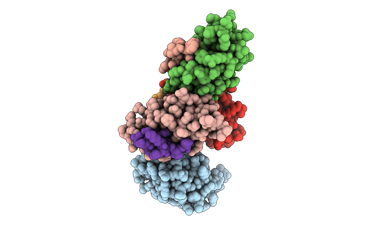
Title:
Structure of the human Mdmx protein bound to the p53 tumor suppressor transactivation domain
Biological Source:
Source Organism(s):
Homo sapiens (Taxon ID: 9606)
Expression System(s):
Method Details:
Experimental Method:
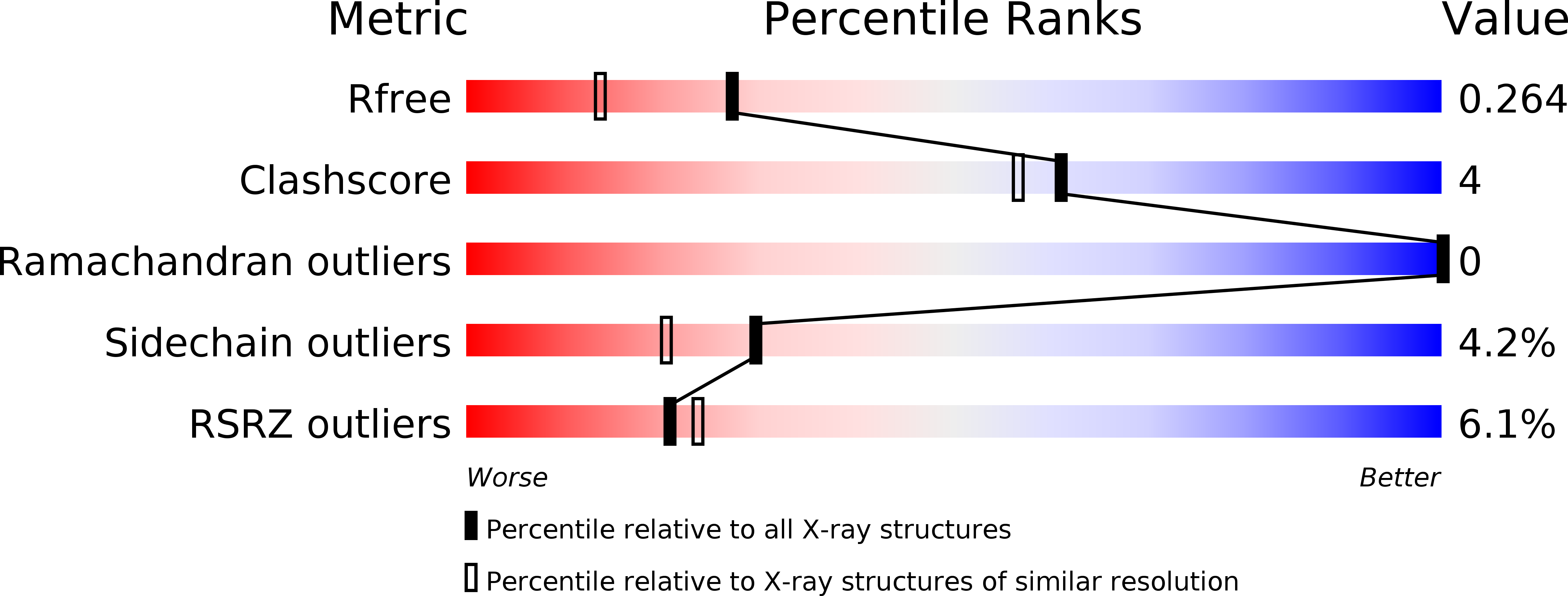
Resolution:
1.90 Å
R-Value Free:
0.25
R-Value Work:
0.19
R-Value Observed:
0.19
Space Group:
P 1 21 1