3DA9 image
Deposition Date 2008-05-29
Release Date 2009-05-19
Last Version Date 2024-11-13
Entry Detail
PDB ID:
3DA9
Keywords:
Title:
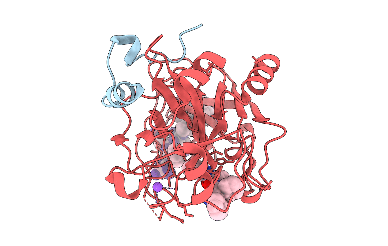
Crystal structure of thrombin in complex with inhibitor
Biological Source:
Source Organism:
Homo sapiens (Taxon ID: 9606)
Method Details:
Experimental Method:
Resolution:
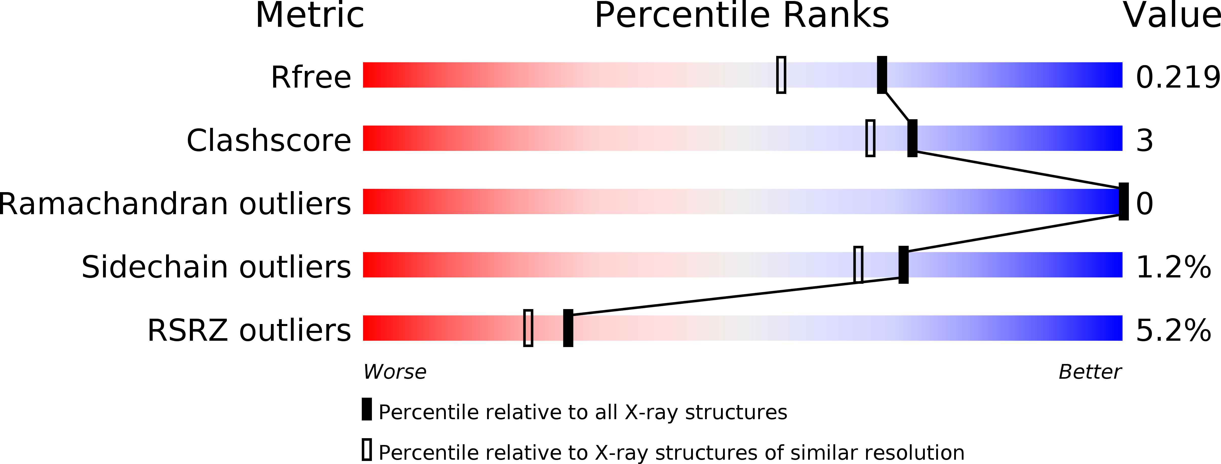
1.80 Å
R-Value Free:
0.21
R-Value Work:
0.18
R-Value Observed:
0.18
Space Group:
C 1 2 1
Macromolecular Entities
Polymer Type:polypeptide(L)
Molecule:Thrombin light chain
Gene (Uniprot):F2
Chain IDs:A
Chain Length:36
Number of Molecules:1
Biological Source:Homo sapiens
Polymer Type:polypeptide(L)
Molecule:Thrombin heavy chain
Gene (Uniprot):F2
Chain IDs:B
Chain Length:259
Number of Molecules:1
Biological Source:Homo sapiens
Modified Residue
Compound ID Chain ID Parent Comp ID Details 2D Image
PTR C TYR O-PHOSPHOTYROSINE
Primary Citation
Compounds binding to the S2-S3 pockets of thrombin.
J.Med.Chem. 52 2708 2715 (2009)
PMID: 19371038 DOI: 10.1021/jm8011849

Abstact

A set of compounds designed to bind to the S2-S3 pockets of thrombin was prepared. These compounds included examples with no interactions in the S1 pocket. Proline, a common P2 in many thrombin inhibitors, was combined with known P3 residues and P1 substituents of varying size and lipophilicity. Binding constants were determined using surface plasmon resonance (SPR) biosensor technology and were found to be in good agreement with results from an enzyme assay. A dramatic increase in affinity (100-1000 times) was seen for compounds incorporating an amino group capable of forming a hydrogen bond with gly216 in the protein backbone. The ligand efficiency was increased by including substituents that form stronger hydrophobic interactions with the P1 pocket. The binding mode was confirmed by X-ray analysis, which revealed the anticipated binding motif that included hydrogen bonds as well as a tightly bound water molecule. A QSAR model indicated that hydrogen bonding and lipophilicity were important for the prediction of binding constants. The results described here may have implications for how directed compound libraries for shallow protein pockets, like S2 and S3 in serine proteases, can be designed.

Legend

Protein

Chemical

Disease

Primary Citation of related structures