Deposition Date
2008-05-21
Release Date
2008-08-12
Last Version Date
2023-08-30
Entry Detail
PDB ID:
3D7P
Keywords:
Title:
Crystal structure of human Transthyretin (TTR) at pH 4.0
Biological Source:
Source Organism(s):
Homo sapiens (Taxon ID: 9606)
Expression System(s):
Method Details:
Experimental Method:
Resolution:
1.72 Å
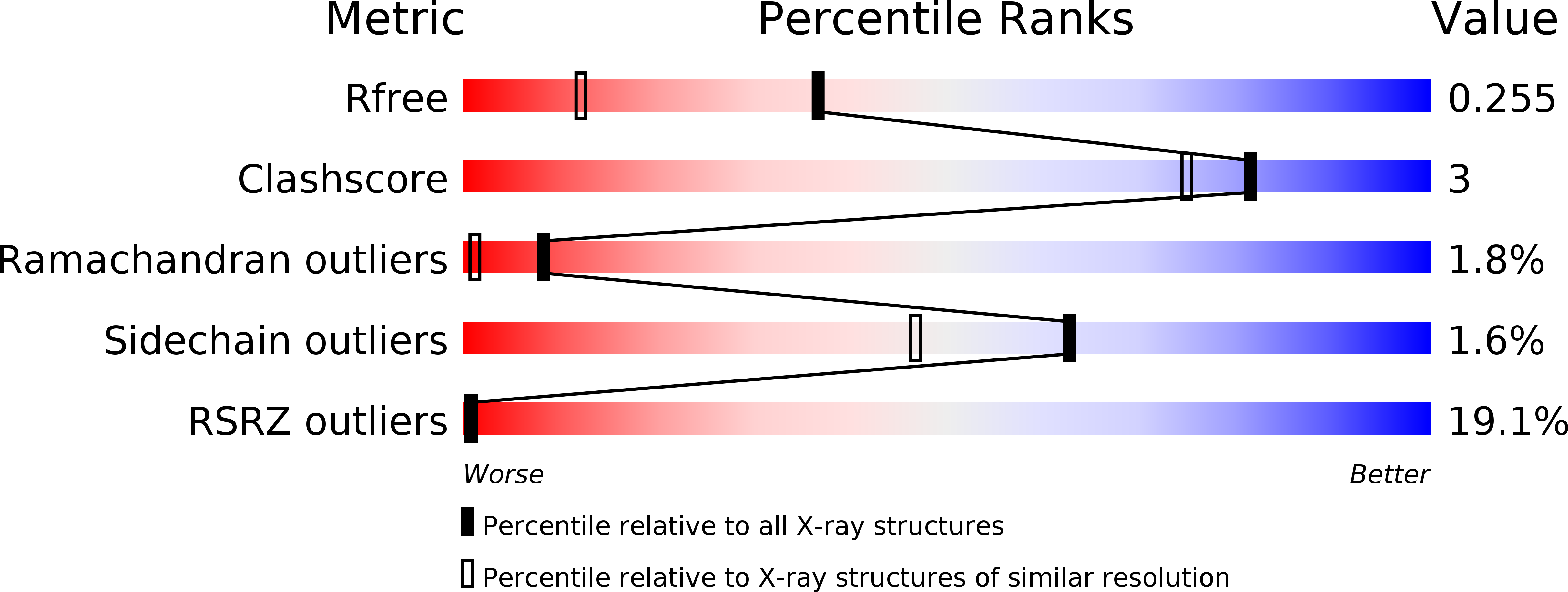
R-Value Free:
0.24
R-Value Work:
0.22
R-Value Observed:
0.22
Space Group:
P 21 21 2