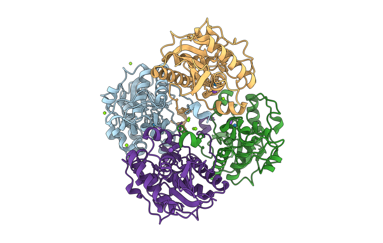
3D6A image
Entry Detail
PDB ID:
3D6A
Keywords:
Title:
Crystal structure of the 2H-phosphatase domain of Sts-2 in complex with tungstate.
Biological Source:
Source Organism:
Host Organism:
PDB Version:
Deposition Date:
2008-05-19
Release Date:
2009-03-03
Method Details:
Experimental Method:
Resolution:
2.25 Å
R-Value Free:
0.25
R-Value Work:
0.20
R-Value Observed:
0.21
Space Group:
P 21 21 21
Macromolecular Entities
Polymer Type:polypeptide(L)
Description:Sts-2 protein
Chain IDs:A, B, C, D
Chain Length:273
Number of Molecules:4
Biological Source:Mus musculus
Primary Citation
Structural and functional characterization of the 2H-phosphatase domain of Sts-2 reveals an acid-dependent phosphatase activity.
Biochemistry 48 1681 1690 (2009)
PMID: 19196006 DOI: 10.1021/bi802219n

Abstact

The suppressors of T cell receptor (TCR) signaling 1 and 2 (Sts-1 and -2, respectively) are multidomain proteins that negatively regulate the signaling of membrane-bound receptors, including TCR and the epidermal growth factor receptor (EGFR). Sts-1 was recently shown to be a new type of protein tyrosine phosphatase (PTP), with the phosphatase activity located within its C-terminal phosphoglycerate mutase (PGM) homology domain and key for the regulation of TCR signaling in T cells. The activity of the related Sts-2 enzyme is significantly less than that of Sts-1. Here we investigate the phosphatase activity of the PGM domain of Sts-2, Sts-2(PGM). The crystal structure of Sts-2(PGM) is remarkably similar to Sts-1(PGM), including conservation of all catalytic residues. Insight into mechanistic details is provided by the structures of the apo, tungstate-bound, and phosphate-bound enzyme. The active site shows stringent specificity, with the k(cat) optimum at pH 5.0 suggesting that Sts-2 might function as an acid-dependent phosphatase. Mutation of active site residues Gln372, Ala446, Glu481, Ser552, and Ser582 to their equivalents in Sts-1 increases the phosphatase activity of Sts-2(PGM) toward model substrates. Overall, our data demonstrate that Sts-2(PGM) adopts the conformation of an active phosphatase whose activity is fundamentally different from that of Sts-1 despite the strong structural homology. They also demonstrate that nonconserved active site residues are responsible for the difference in activity between the two isoforms. These differences reflect possible distinct physiological substrates.

Legend

Protein

Chemical

Disease

Primary Citation of related structures