3D5G image
Deposition Date 2008-05-16
Release Date 2009-05-26
Last Version Date 2024-10-16
Entry Detail
PDB ID:
3D5G
Keywords:
Title:
Structure of ribonuclease Sa2 complexes with mononucleotides: new aspects of catalytic reaction and substrate recognition
Biological Source:
Source Organism:
Host Organism:
Method Details:
Experimental Method:
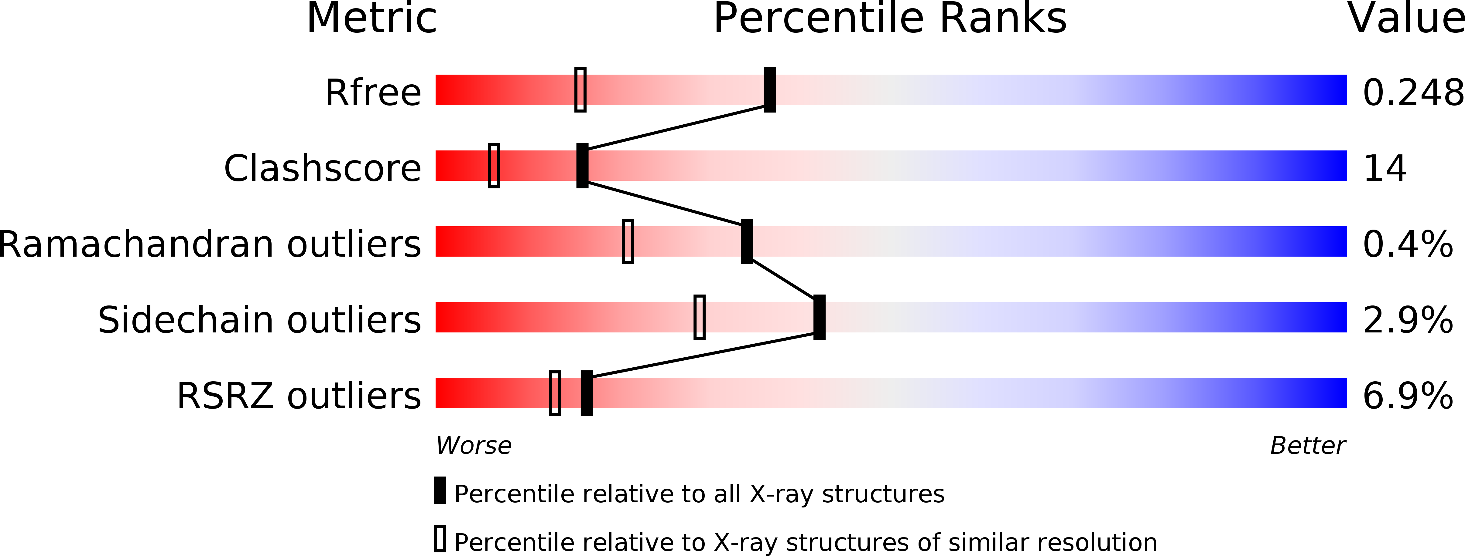
Resolution:
1.80 Å
R-Value Free:
0.24
R-Value Work:
0.17
R-Value Observed:
0.17
Space Group:
C 1 2 1
Macromolecular Entities
Polymer Type:polypeptide(L)
Molecule:Ribonuclease
Gene (Uniprot):ribonuclease
Chain IDs:A, B, C
Chain Length:97
Number of Molecules:3
Biological Source:Streptomyces aureofaciens
Ligand Molecules
Primary Citation
Structure of RNase Sa2 complexes with mononucleotides - new aspects of catalytic reaction and substrate recognition
Febs J. 276 4156 4168 (2009)
PMID: 19558492 DOI: 10.1111/j.1742-4658.2009.07125.x

Abstact

Although the mechanism of RNA cleavage by RNases has been studied for many years, there remain aspects that have not yet been fully clarified. We have solved the crystal structures of RNase Sa2 in the apo form and in complexes with mononucleotides. These structures provide more details about the mechanism of RNA cleavage by RNase Sa2. In addition to Glu56 and His86, which are the principal catalytic residues, an important role in the first reaction step of RNA cleavage also seems to be played by Arg67 and Arg71, which are located in the phosphate-binding site and form hydrogen bonds with the oxygens of the phosphate group of the mononucleotides. Their positive charge very likely causes polarization of the bonds between the oxygens and the phosphorus atom, leading to electron deficiency on the phosphorus atom and facilitating nucleophilic attack by O2' of the ribose on the phosphorus atom, leading to cyclophosphate formation. The negatively charged Glu56 is in position to attract the proton from O2' of the ribose. Extended molecular docking of mononucleotides, dinucleotides and trinucleotides into the active site of the enzyme allowed us to better understand the guanosine specificity of RNase Sa2 and to predict possible binding subsites for the downstream base and ribose of the second and third nucleotides.

Legend

Protein

Chemical

Disease

Primary Citation of related structures