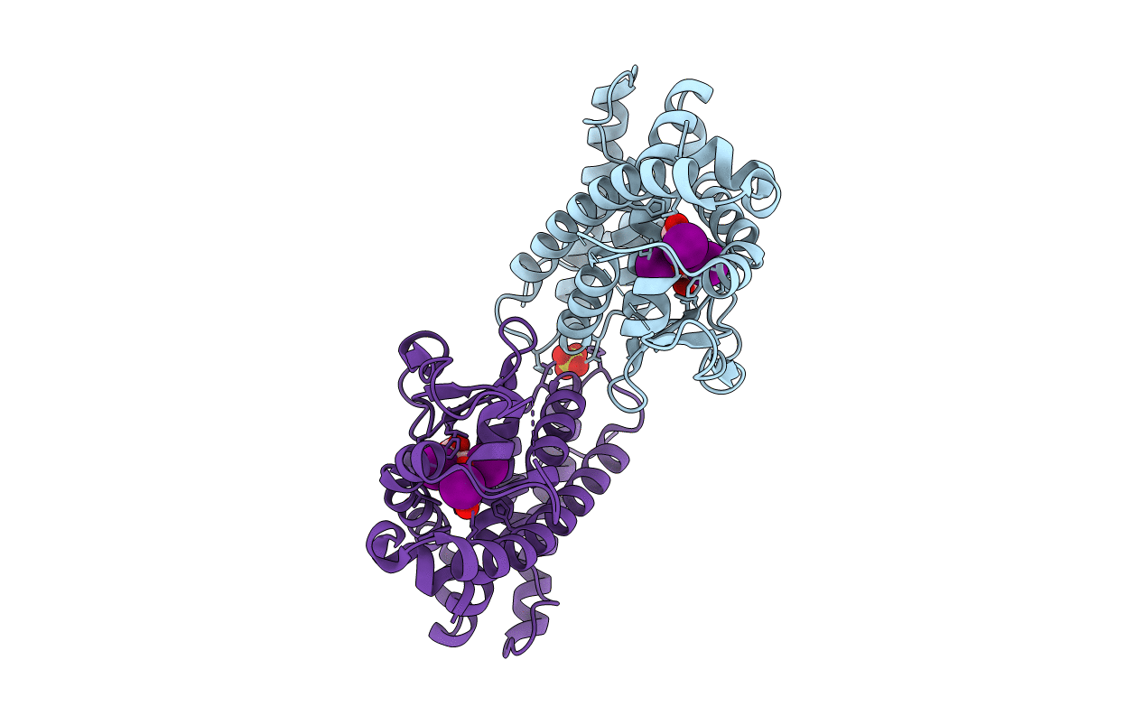
3D57 image
Deposition Date 2008-05-15
Release Date 2008-10-14
Last Version Date 2023-08-30
Entry Detail
PDB ID:
3D57
Title:
TR Variant D355R
Biological Source:
Source Organism:
Homo sapiens (Taxon ID: 9606)
Host Organism:
Method Details:
Experimental Method:
Resolution:
2.20 Å
R-Value Free:
0.24
R-Value Work:
0.21
R-Value Observed:
0.21
Space Group:
P 1 21 1
Macromolecular Entities
Polymer Type:polypeptide(L)
Molecule:Thyroid hormone receptor beta
Gene (Uniprot):THRB
Mutations:D355R
Chain IDs:A, B
Chain Length:266
Number of Molecules:2
Biological Source:Homo sapiens
Primary Citation
Molecular basis for dimer formation of TRbeta variant D355R.
Proteins 75 111 117 (2008)
PMID: 18798561 DOI: 10.1002/prot.22225

Abstact

Protein quality and stability are critical during protein purification for X-ray crystallography. A target protein that is easy to manipulate and crystallize becomes a valuable product useful for high-throughput crystallography for drug design and discovery. In this work, a single surface mutation, D355R, was shown to be crucial for converting the modestly stable monomeric ligand binding domain of the human thyroid hormone receptor (TR LBD) into a stable dimer. The structure of D335R TR LBD mutant was solved using X-ray crystallography and refined to 2.2 A resolution with R(free)/R values of 24.5/21.7. The crystal asymmetric unit reveals the TR dimer with two molecules of the hormone-bound LBD related by twofold symmetry. The ionic interface between the two LBDs comprises residues within loop H10-H11 and loop H6-H7 as well as the C-terminal halves of helices 8 of both protomers. Direct intermolecular contacts formed between the introduced residue Arg 355 of one TR molecule and Glu 324 of the second molecule become a part of the extended dimerization interface of 1330 A(2) characteristic for a strong complex assembly that is additionally strengthened by buffer solutes.

Legend

Protein

Chemical

Disease

Primary Citation of related structures