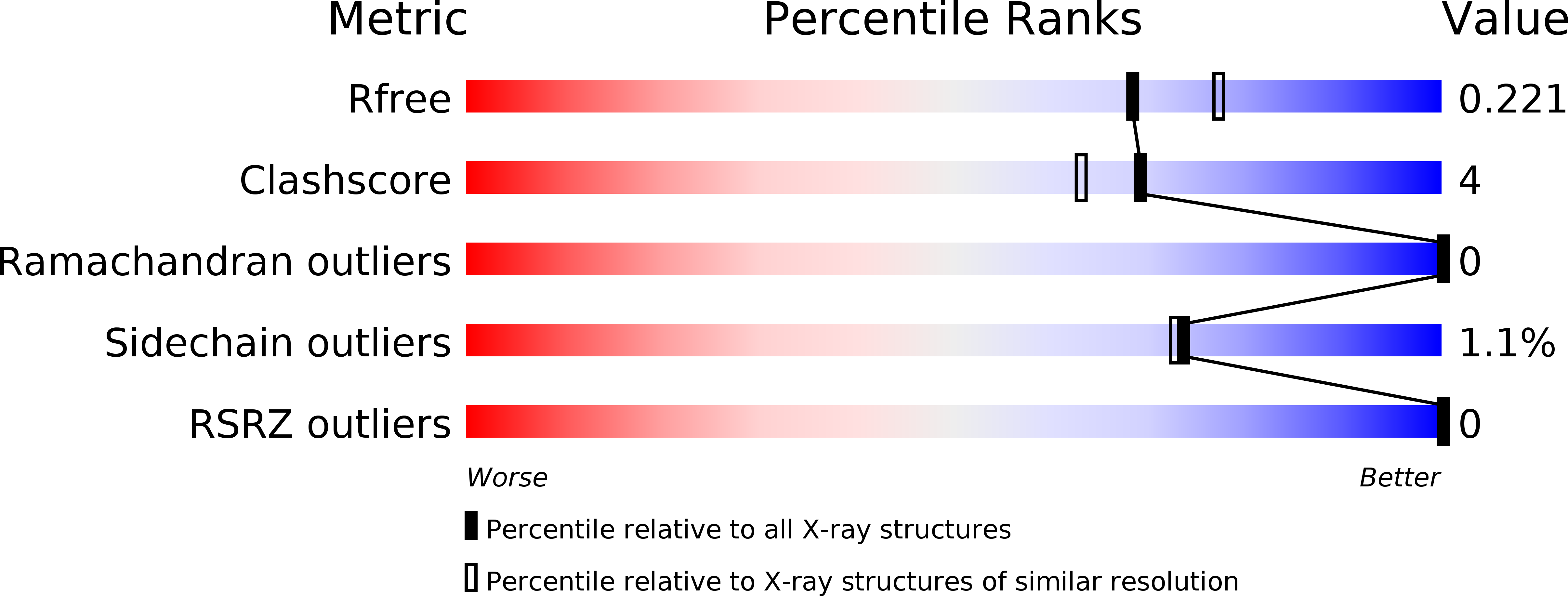
3D4M image
Deposition Date 2008-05-14
Release Date 2008-10-28
Last Version Date 2024-10-16
Entry Detail
PDB ID:
3D4M
Keywords:
Title:
Glutaredoxin 2 oxidized structure
Biological Source:
Source Organism:
Host Organism:
Method Details:
Experimental Method:
Resolution:
2.05 Å
R-Value Free:
0.21
R-Value Work:
0.18
R-Value Observed:
0.18
Space Group:
P 41 21 2
Macromolecular Entities
Polymer Type:polypeptide(L)
Molecule:Glutaredoxin-2, mitochondrial
Gene (Uniprot):GRX2
Chain IDs:A
Chain Length:109
Number of Molecules:1
Biological Source:Saccharomyces cerevisiae
Primary Citation
Structural aspects of the distinct biochemical properties of glutaredoxin 1 and glutaredoxin 2 from Saccharomyces cerevisiae.
J.Mol.Biol. 385 889 901 (2009)
PMID: 18992757 DOI: 10.1016/j.jmb.2008.10.055

Abstact

Glutaredoxins (Grxs) are small (9-12 kDa) heat-stable proteins that are ubiquitously distributed. In Saccharomyces cerevisiae, seven Grx enzymes have been identified. Two of them (yGrx1 and yGrx2) are dithiolic, possessing a conserved Cys-Pro-Tyr-Cys motif. Here, we show that yGrx2 has a specific activity 15 times higher than that of yGrx1, although these two oxidoreductases share 64% identity and 85% similarity with respect to their amino acid sequences. Further characterization of the enzymatic activities through two-substrate kinetics analysis revealed that yGrx2 possesses a lower K(M) for glutathione and a higher turnover than yGrx1. To better comprehend these biochemical differences, the pK(a) of the N-terminal active-site cysteines (Cys27) of these two proteins and of the yGrx2-C30S mutant were determined. Since the pK(a) values of the yGrx1 and yGrx2 Cys27 residues are very similar, these parameters cannot account for the difference observed between their specific activities. Therefore, crystal structures of yGrx2 in the oxidized form and with a glutathionyl mixed disulfide were determined at resolutions of 2.05 and 1.91 A, respectively. Comparisons of yGrx2 structures with the recently determined structures of yGrx1 provided insights into their remarkable functional divergence. We hypothesize that the substitutions of Ser23 and Gln52 in yGrx1 by Ala23 and Glu52 in yGrx2 modify the capability of the active-site C-terminal cysteine to attack the mixed disulfide between the N-terminal active-site cysteine and the glutathione molecule. Mutagenesis studies supported this hypothesis. The observed structural and functional differences between yGrx1 and yGrx2 may reflect variations in substrate specificity.

Legend

Protein

Chemical

Disease

Primary Citation of related structures