3D41 image
Entry Detail
PDB ID:
3D41
Keywords:
Title:
Crystal structure of fosfomycin resistance kinase FomA from Streptomyces wedmorensis complexed with MgAMPPNP and fosfomycin
Biological Source:
Source Organism:
Host Organism:
PDB Version:
Deposition Date:
2008-05-13
Release Date:
2008-08-12
Method Details:
Experimental Method:
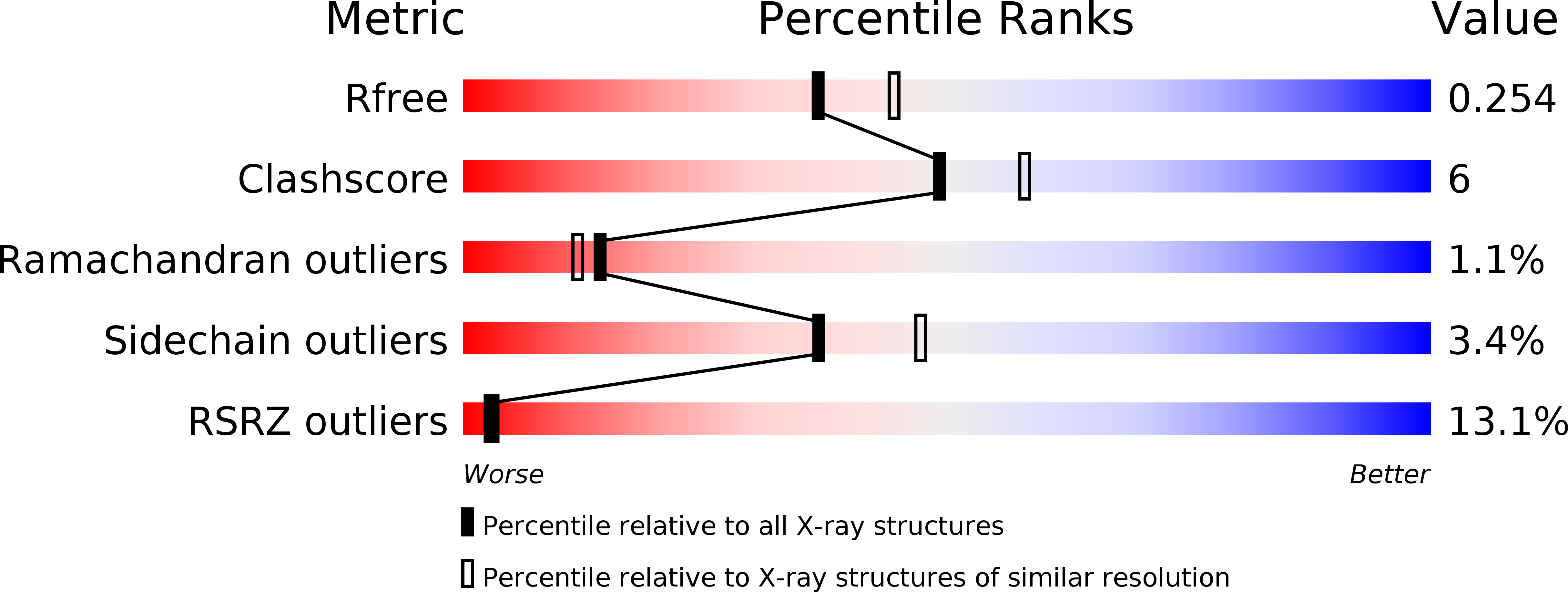
Resolution:
2.20 Å
R-Value Free:
0.25
R-Value Work:
0.19
R-Value Observed:
0.19
Space Group:
P 32 2 1
Macromolecular Entities
Polymer Type:polypeptide(L)
Description:FomA protein
Chain IDs:A
Chain Length:286
Number of Molecules:1
Biological Source:Streptomyces wedmorensis
Primary Citation
Crystal Structure of Fosfomycin Resistance Kinase FomA from Streptomyces wedmorensis.
J.Biol.Chem. 283 28518 28526 (2008)
PMID: 18701452 DOI: 10.1074/jbc.M803709200

Abstact

The fosfomycin resistance protein FomA inactivates fosfomycin by phosphorylation of the phosphonate group of the antibiotic in the presence of ATP and Mg(II). We report the crystal structure of FomA from the fosfomycin biosynthetic gene cluster of Streptomyces wedmorensis in complex with diphosphate and in ternary complex with the nonhydrolyzable ATP analog adenosine 5'-(beta,gamma-imido)-triphosphate (AMPPNP), Mg(II), and fosfomycin, at 1.53 and 2.2 angstroms resolution, respectively. The polypeptide exhibits an open alphabetaalpha sandwich fold characteristic for the amino acid kinase family of enzymes. The diphosphate complex shows significant disorder in loops surrounding the active site. As a result, the nucleotide-binding site is wide open. Binding of the substrates is followed by the partial closure of the active site and ordering of the alpha2-helix. Structural comparison with N-acetyl-L-glutamate kinase shows several similarities in the site of phosphoryl transfer: 1) preservation of architecture of the catalytical amino acids of N-acetyl-L-glutamate kinase (Lys9, Lys216, and Asp150 in FomA); 2) good superposition of the phosphate acceptor groups of the substrates, and 3) good superposition of the diphosphate molecule with the beta- and gamma-phosphates of AMPPNP, suggesting that the reaction could proceed by an associative in-line mechanism. However, differences in conformations of the triphosphate moiety of AMPPNP molecules, the long distance (5.1 angstroms) between the phosphate acceptor and donor groups in FomA, and involvement of Lys18 instead of Lys9 in binding with the gamma-phosphate may indicate a different reaction mechanism. The present work identifies the active site residues of FomA responsible for substrate binding and specificity and proposes their roles in catalysis.

Legend

Protein

Chemical

Disease

Primary Citation of related structures