3D3T image
Deposition Date 2008-05-12
Release Date 2008-07-08
Last Version Date 2024-02-21
Entry Detail
PDB ID:
3D3T
Keywords:
Title:
Crystal Structure of HIV-1 CRF01_AE in complex with the substrate p1-p6
Biological Source:
Source Organism:
Host Organism:
Method Details:
Experimental Method:
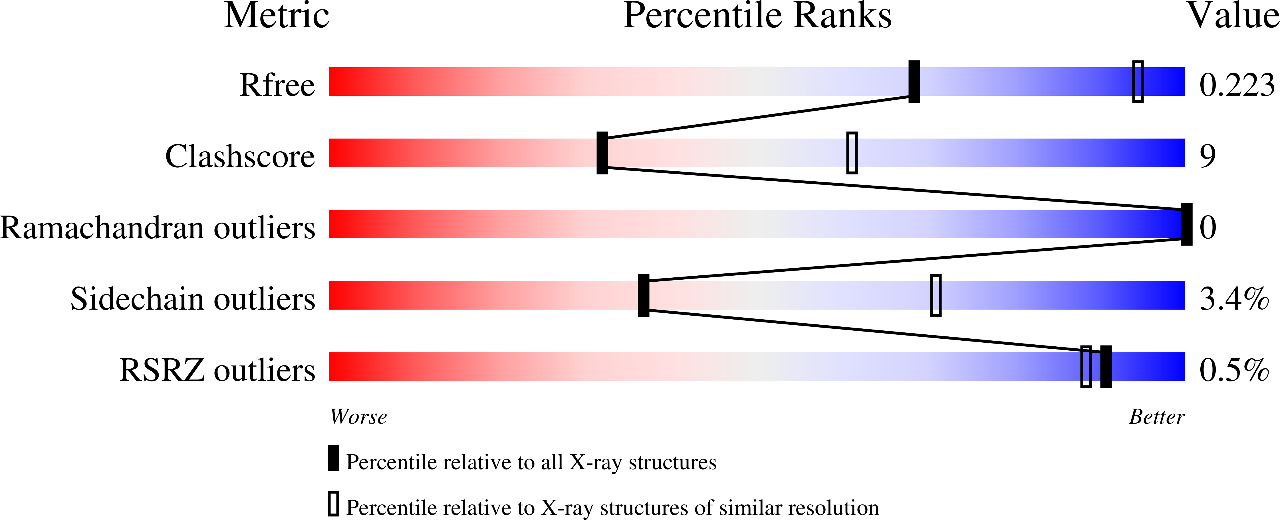
Resolution:
2.80 Å
R-Value Free:
0.25
R-Value Work:
0.19
R-Value Observed:
0.20
Space Group:
P 61
Macromolecular Entities
Polymer Type:polypeptide(L)
Molecule:HIV-1 protease
Gene (Uniprot):gag-pol
Mutations:D25N
Chain IDs:A, B
Chain Length:99
Number of Molecules:2
Biological Source:Human immunodeficiency virus 1
Polymer Type:polypeptide(L)
Molecule:P1-P6 SUBSTRATE PEPTIDE
Gene (Uniprot):gag
Chain IDs:C (auth: P)
Chain Length:10
Number of Molecules:1
Biological Source:
Primary Citation
Structural analysis of human immunodeficiency virus type 1 CRF01_AE protease in complex with the substrate p1-p6.
J.Virol. 82 6762 6766 (2008)
PMID: 18434392 DOI: 10.1128/JVI.00018-08

Abstact

The effect of amino acid variability between human immunodeficiency virus type 1 (HIV-1) clades on structure and the emergence of resistance mutations in HIV-1 protease has become an area of significant interest in recent years. We determined the first crystal structure of the HIV-1 CRF01_AE protease in complex with the p1-p6 substrate to a resolution of 2.8 A. Hydrogen bonding between the flap hinge and the protease core regions shows significant structural rearrangements in CRF01_AE protease compared to the clade B protease structure.

Legend

Protein

Chemical

Disease

Primary Citation of related structures