3D3K image
Deposition Date 2008-05-12
Release Date 2008-08-26
Last Version Date 2024-03-20
Entry Detail
PDB ID:
3D3K
Keywords:
Title:
Crystal structure of human Edc3p
Biological Source:
Source Organism:
Homo sapiens (Taxon ID: 9606)
Host Organism:
Method Details:
Experimental Method:
Resolution:
2.20 Å
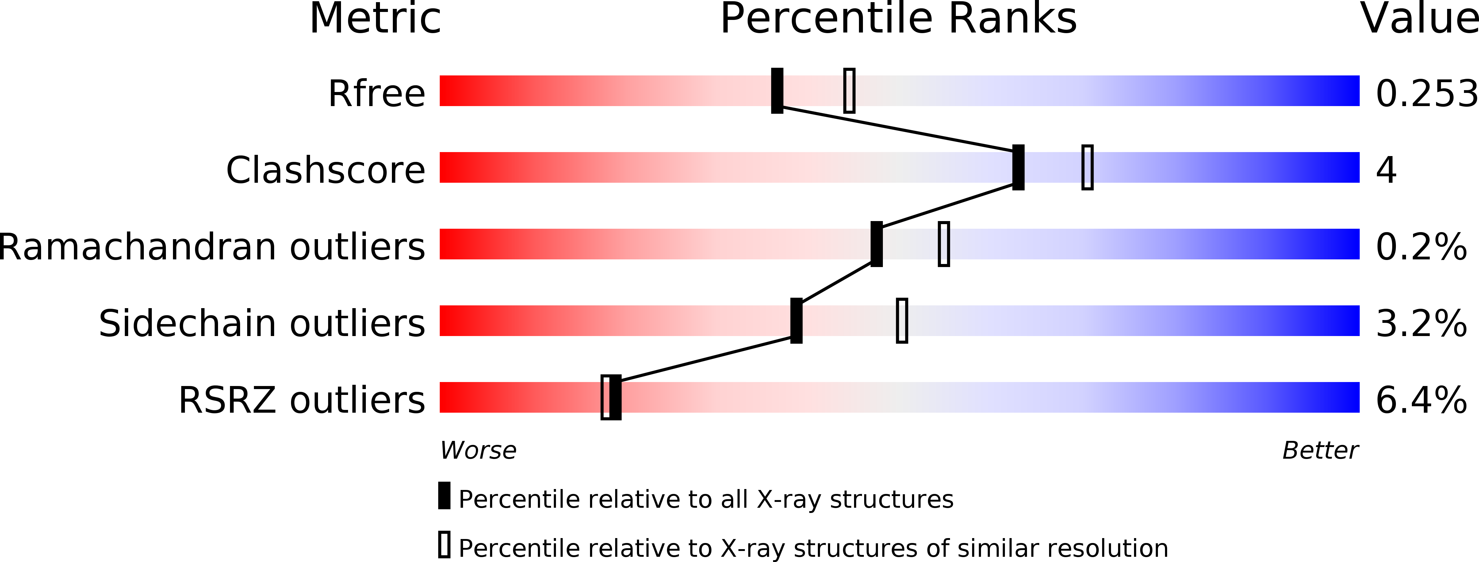
R-Value Free:
0.25
R-Value Work:
0.22
R-Value Observed:
0.22
Space Group:
C 2 2 21
Macromolecular Entities
Structures with similar UniProt ID
Protein Blast
Polymer Type:polypeptide(L)
Molecule:Enhancer of mRNA-decapping protein 3
Gene (Uniprot):EDC3
Chain IDs:A, B, C, D
Chain Length:259
Number of Molecules:4
Biological Source:Homo sapiens
Primary Citation
Crystal structure of human Edc3 and its functional implications
Mol.Cell.Biol. 28 5965 5976 (2008)
PMID: 18678652 DOI: 10.1128/MCB.00761-08

Abstact

Edc3 is an enhancer of decapping and serves as a scaffold that aggregates mRNA ribonucleoproteins together for P-body formation. Edc3 forms a network of interactions with the components of the mRNA decapping machinery and has a modular domain architecture consisting of an N-terminal Lsm domain, a central FDF domain, and a C-terminal YjeF-N domain. We have determined the crystal structure of the N-terminally truncated human Edc3 at a resolution of 2.2 A. The structure reveals that the YjeF-N domain of Edc3 possesses a divergent Rossmann fold topology that forms a dimer, which is supported by sedimentation velocity and sedimentation equilibrium analysis in solution. The dimerization interface of Edc3 is highly conserved in eukaryotes despite the overall low sequence homology across species. Structure-based site-directed mutagenesis revealed dimerization is required for efficient RNA binding, P-body formation, and likely for regulating the yeast Rps28B mRNA as well, suggesting that the dimeric form of Edc3 is a structural and functional unit in mRNA degradation.

Legend

Protein

Chemical

Disease

Primary Citation of related structures