3D2Z image
Deposition Date 2008-05-09
Release Date 2009-06-16
Last Version Date 2024-10-30
Entry Detail
PDB ID:
3D2Z
Keywords:
Title:
Complex of the N-acetylmuramyl-L-alanine amidase AmiD from E.coli with the product L-Ala-D-gamma-Glu-L-Lys
Biological Source:
Host Organism:
Method Details:
Experimental Method:
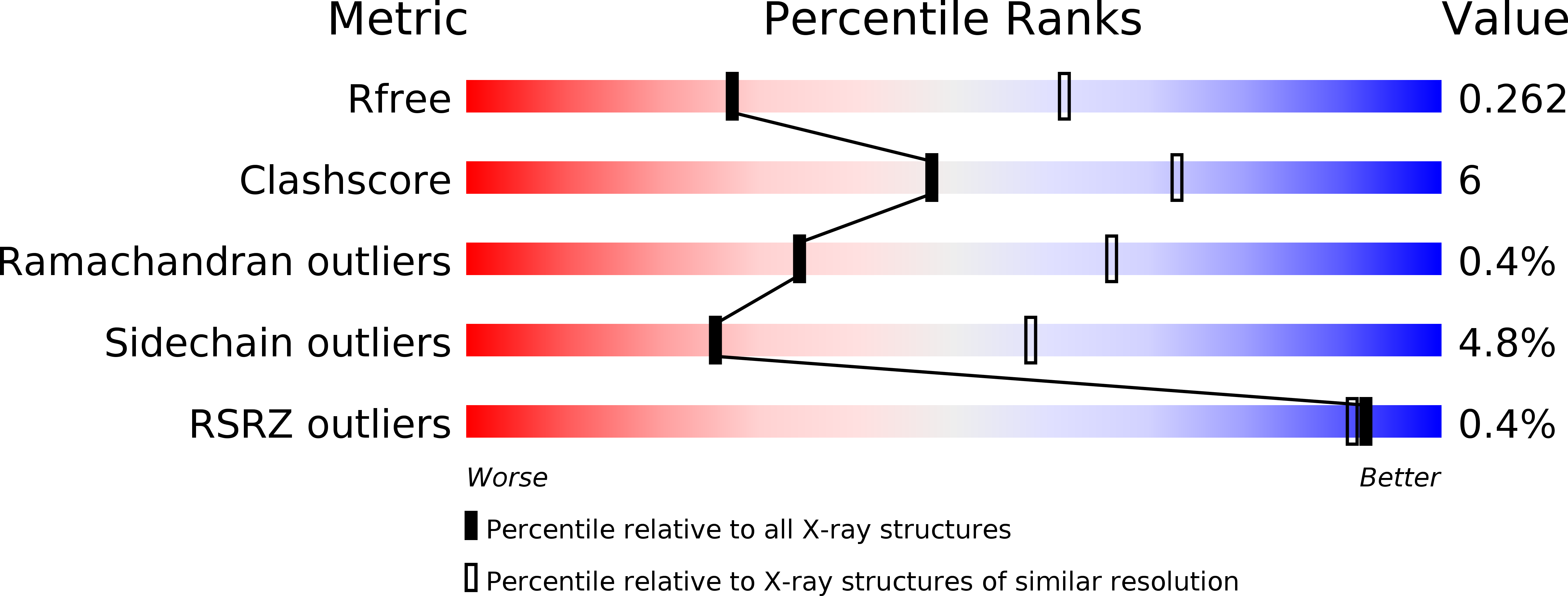
Resolution:
2.80 Å
R-Value Free:
0.26
R-Value Work:
0.20
R-Value Observed:
0.20
Space Group:
P 61 2 2
Macromolecular Entities
Structures with similar UniProt ID
Protein Blast
Polymer Type:polypeptide(L)
Molecule:N-acetylmuramoyl-L-alanine amidase amiD
Gene (Uniprot):amiD
Chain IDs:A
Chain Length:261
Number of Molecules:1
Biological Source:Escherichia coli str. K12 substr. MG1655
Polymer Type:polypeptide(L)
Molecule:L-Ala-D-gamma-Glu-L-Lys peptide
Chain IDs:B
Chain Length:3
Number of Molecules:1
Biological Source:
Primary Citation
Specific Structural Features of the N-Acetylmuramoyl-l-Alanine Amidase AmiD from Escherichia coli and Mechanistic Implications for Enzymes of This Family.
J.Mol.Biol. 397 249 259 (2010)
PMID: 20036252 DOI: 10.1016/j.jmb.2009.12.038

Abstact

AmiD is the fifth identified N-acetylmuramoyl-L-alanine zinc amidase of Escherichia coli. This periplasmic lipoprotein is anchored in the outer membrane and has a broad specificity. AmiD is capable of cleaving the intact peptidoglycan (PG) as well as soluble fragments containing N-acetylmuramic acid regardless of the presence of an anhydro form or not, unlike the four other amidases, AmiA, AmiB, AmiC, and AmpD, which have some specificity. AmiD function is, however, not clearly established but it could be part of the enzymatic machinery involved in the PG turnover in E. coli. We solved three structures of the E. coli zinc amidase AmiD devoid of its lipidic anchorage: the holoenzyme, the apoenzyme in complex with the substrate anhydro-N-acetylmuramic-acid-L-Ala-gamma-d-Glu-L-Lys, and the holoenzyme in complex with the L-Ala-gamma-D-Glu-L-Lys peptide, the product of the hydrolysis of this substrate by AmiD. The AmiD structure shows a relatively flexible N-terminal extension that allows an easy reach of the PG by the enzyme inserted into the outer membrane. The C-terminal domain provides a potential extended geometrical complementarity to the substrate. AmiD shares a common fold with AmpD, the bacteriophage T7 lysozyme, and the PG recognition proteins, which are receptor proteins involved in the innate immune responses of a wide range of organisms. Analysis of the different structures reveals the similarity between the catalytic mechanism of zinc amidases of the AmiD family and the thermolysin-related zinc peptidases.

Legend

Protein

Chemical

Disease

Primary Citation of related structures