
Deposition Date
2008-05-08
Release Date
2008-12-23
Last Version Date
2023-08-30
Entry Detail
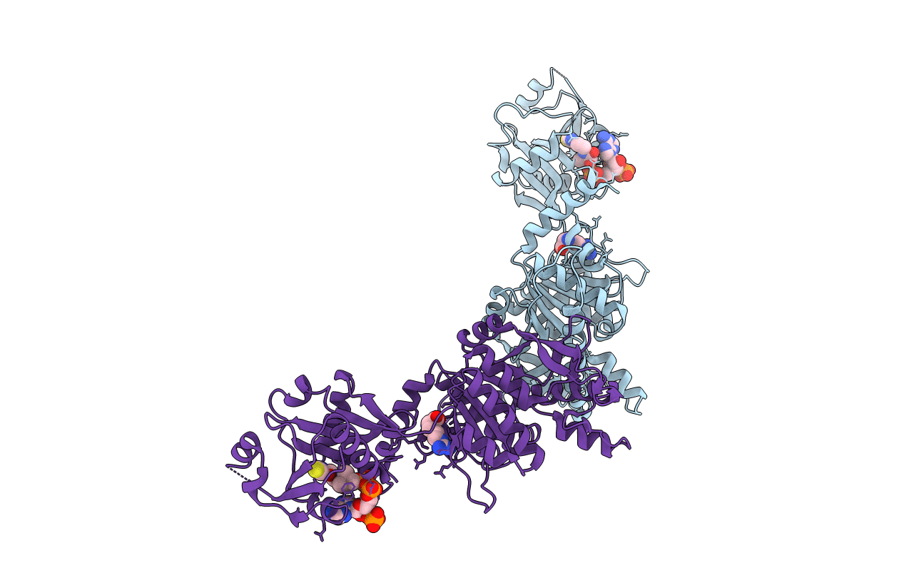
PDB ID:
3D2P
Keywords:
Title:
Crystal structure of N-acetylglutamate synthase from Neisseria gonorrhoeae complexed with coenzyme A and L-arginine
Biological Source:
Source Organism(s):
Neisseria gonorrhoeae (Taxon ID: 485)
Expression System(s):
Method Details:
Experimental Method:
Resolution:
2.56 Å
R-Value Free:
0.27
R-Value Work:
0.21
R-Value Observed:
0.21
Space Group:
P 3 1 2
