3D1F image
Deposition Date 2008-05-05
Release Date 2008-07-29
Last Version Date 2023-08-30
Entry Detail
PDB ID:
3D1F
Title:
Crystal structure of E. coli sliding clamp (beta) bound to a polymerase III peptide
Biological Source:
Source Organism:
Escherichia coli (Taxon ID: )
Host Organism:
Method Details:
Experimental Method:
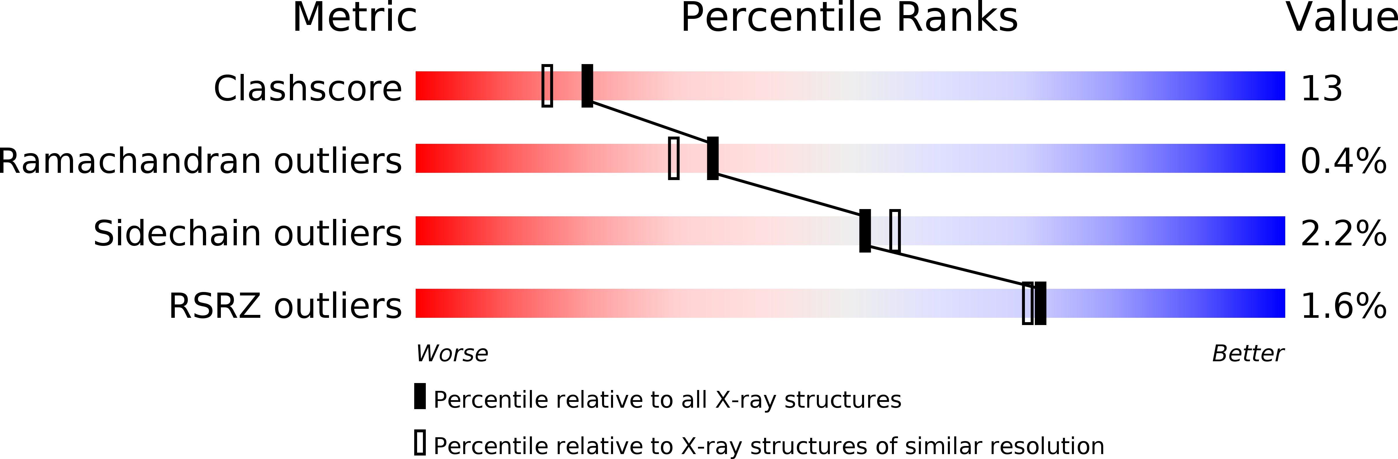
Resolution:
2.00 Å
R-Value Free:
0.25
R-Value Work:
0.21
Space Group:
P 32
Macromolecular Entities
Polymer Type:polypeptide(L)
Molecule:DNA polymerase III subunit beta
Gene (Uniprot):dnaN
Chain IDs:A, B
Chain Length:366
Number of Molecules:2
Biological Source:Escherichia coli
Polymer Type:polypeptide(L)
Molecule:Nonapeptide from polymerase III C-terminal
Chain IDs:C (auth: P), D (auth: Q)
Chain Length:9
Number of Molecules:2
Biological Source:
Primary Citation
Structure of a small-molecule inhibitor of a DNA polymerase sliding clamp.
Proc.Natl.Acad.Sci.Usa 105 11116 11121 (2008)
PMID: 18678908 DOI: 10.1073/pnas.0804754105

Abstact

DNA polymerases attach to the DNA sliding clamp through a common overlapping binding site. We identify a small-molecule compound that binds the protein-binding site in the Escherichia coli beta-clamp and differentially affects the activity of DNA polymerases II, III, and IV. To understand the molecular basis of this discrimination, the cocrystal structure of the chemical inhibitor is solved in complex with beta and is compared with the structures of Pol II, Pol III, and Pol IV peptides bound to beta. The analysis reveals that the small molecule localizes in a region of the clamp to which the DNA polymerases attach in different ways. The results suggest that the small molecule may be useful in the future to probe polymerase function with beta, and that the beta-clamp may represent an antibiotic target.

Legend

Protein

Chemical

Disease

Primary Citation of related structures