3D17 image
Deposition Date 2008-05-05
Release Date 2008-06-03
Last Version Date 2025-11-12
Entry Detail
PDB ID:
3D17
Keywords:
Title:
A triply ligated crystal structure of relaxed state human hemoglobin
Biological Source:
Source Organism(s):
Homo sapiens (Taxon ID: )
Method Details:
Experimental Method:
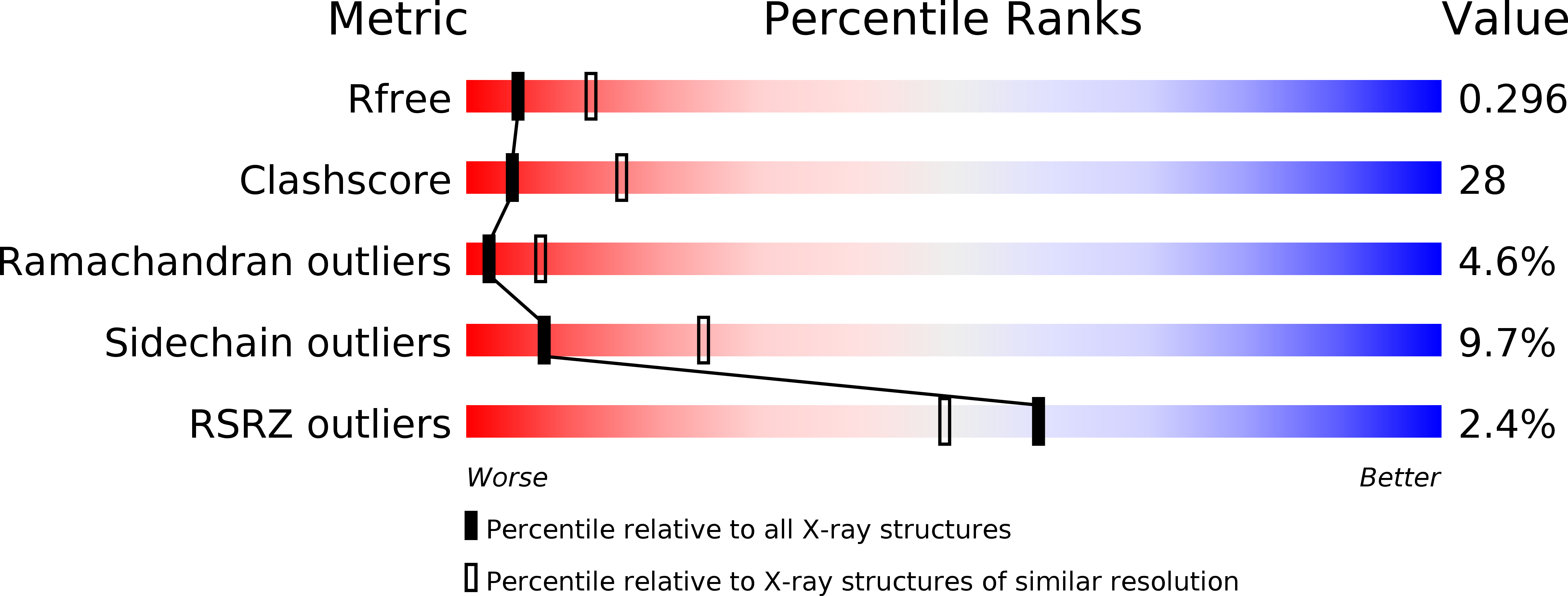
Resolution:
2.80 Å
R-Value Free:
0.29
R-Value Work:
0.23
R-Value Observed:
0.25
Space Group:
P 41 21 2
Macromolecular Entities
Structures with similar UniProt ID
Protein Blast
Polymer Type:polypeptide(L)
Molecule:Hemoglobin subunit alpha
Gene (Uniprot):HBA1, HBA2
Chain IDs:A, C
Chain Length:141
Number of Molecules:2
Biological Source:Homo sapiens
Structures with similar UniProt ID
Protein Blast
Polymer Type:polypeptide(L)
Molecule:Hemoglobin subunit beta
Gene (Uniprot):HBB
Chain IDs:B, D
Chain Length:146
Number of Molecules:2
Biological Source:Homo sapiens
Primary Citation
Structure of relaxed-state human hemoglobin: insight into ligand uptake, transport and release.
Acta Crystallogr.,Sect.D 65 41 48 (2009)
PMID: 19153465 DOI: 10.1107/S0907444908037256

Abstact

Hemoglobin was one of the first protein structures to be determined by X-ray crystallography and served as a basis for the two-state MWC model for the mechanism of allosteric proteins. Since then, there has been an ongoing debate about whether Hb allostery involves the unliganded tense T state and the liganded relaxed R state or whether it involves the T state and an ensemble of liganded relaxed states. In fact, the former model is inconsistent with many functional observations, as well as the recent discoveries of several relaxed-state Hb structures such as RR2, R3 and R2. One school of thought has suggested the R2 state to be the physiologically relevant relaxed end state, with the R state mediating the T-->R2 transition. X-ray studies have been performed on human carbonmonoxy Hb at a resolution of 2.8 A. The ensuing liganded quaternary structure is different from previously reported liganded Hb structures. The distal beta-heme pocket is the largest when compared with other liganded Hb structures, partly owing to rotation of betaHis63(E7) out of the distal pocket, creating a ligand channel to the solvent. The structure also shows unusually smaller alpha- and beta-clefts. Results from this study taken in conjunction with previous findings suggest that multiple liganded Hb states with different quaternary structures may be involved in ligand uptake, stabilization, transport and release.

Legend

Protein

Chemical

Disease

Primary Citation of related structures
Feedback Form
Name
Email
Institute
Feedback