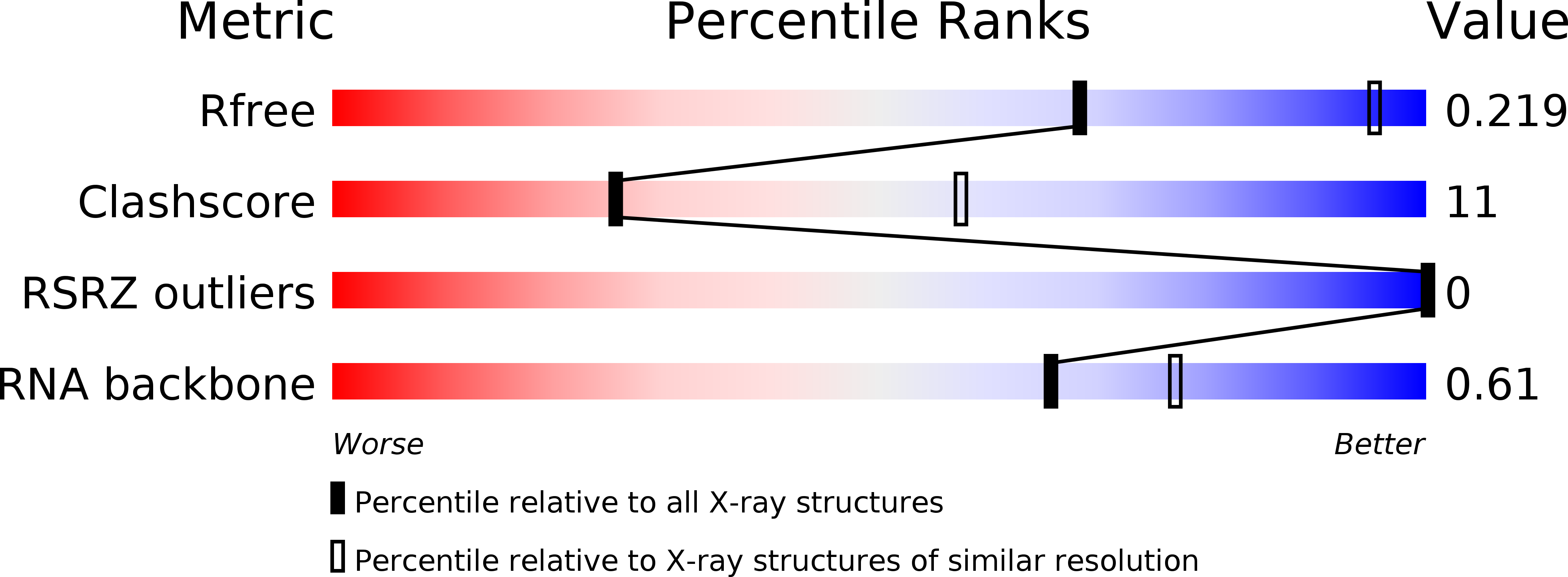
3D0X image
Deposition Date 2008-05-02
Release Date 2008-07-01
Last Version Date 2023-08-30
Entry Detail
PDB ID:
3D0X
Keywords:
Title:
Crystal Structure of the unbound lysine riboswitch
Biological Source:
Source Organism:
(Taxon ID: ) (Taxon ID: )
Method Details:
Experimental Method:
Resolution:
2.95 Å
R-Value Free:
0.22
R-Value Work:
0.18
R-Value Observed:
0.19
Space Group:
P 32
Macromolecular Entities
Polymer Type:polyribonucleotide
Molecule:RNA (161-MER)
Chain IDs:A
Chain Length:161
Number of Molecules:1
Biological Source:
Primary Citation
Crystal structure of the lysine riboswitch regulatory mRNA element.
J.Biol.Chem. 283 22347 22351 (2008)
PMID: 18593706 DOI: 10.1074/jbc.C800120200

Abstact

Riboswitches are metabolite-sensitive elements found in mRNAs that control gene expression through a regulatory secondary structural switch. Along with regulation of lysine biosynthetic genes, mutations within the lysine-responsive riboswitch (L-box) play a role in the acquisition of resistance to antimicrobial lysine analogs. To understand the structural basis for lysine binding, we have determined the 2.8 angstroms resolution crystal structure of lysine bound to the Thermotoga maritima asd lysine riboswitch ligand-binding domain. The structure reveals a complex architecture scaffolding a binding pocket completely enveloping lysine. Mutations conferring antimicrobial resistance cluster around this site as well as highly conserved long range interactions, indicating that they disrupt lysine binding or proper folding of the RNA. Comparison of the free and bound forms by x-ray crystallography, small angle x-ray scattering, and chemical probing reveals almost identical structures, indicating that lysine induces only limited and local conformational changes upon binding.

Legend

Protein

Chemical

Disease

Primary Citation of related structures