Deposition Date
2008-04-26
Release Date
2008-07-08
Last Version Date
2024-02-21
Entry Detail
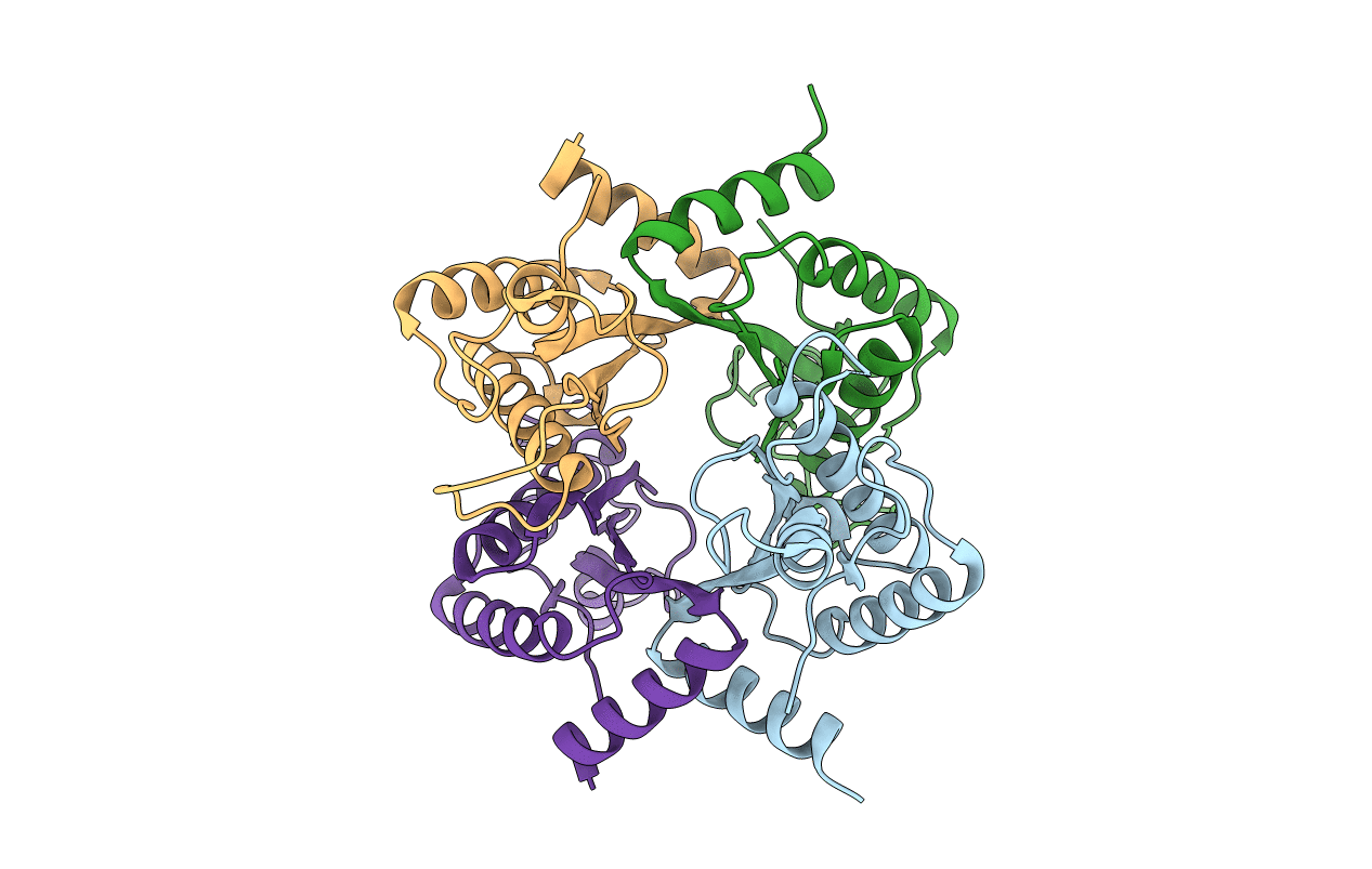
PDB ID:
3CYP
Keywords:
Title:
The crystal structure of the C-terminal domain of Helicobacter pylori MotB (residues 125-256).
Biological Source:
Source Organism(s):
Helicobacter pylori (Taxon ID: )
Expression System(s):
Method Details:
Experimental Method:
Resolution:
1.60 Å
R-Value Free:
0.22
R-Value Work:
0.18
R-Value Observed:
0.18
Space Group:
P 1 21 1