3CXN image
Entry Detail
PDB ID:
3CXN
Keywords:
Title:
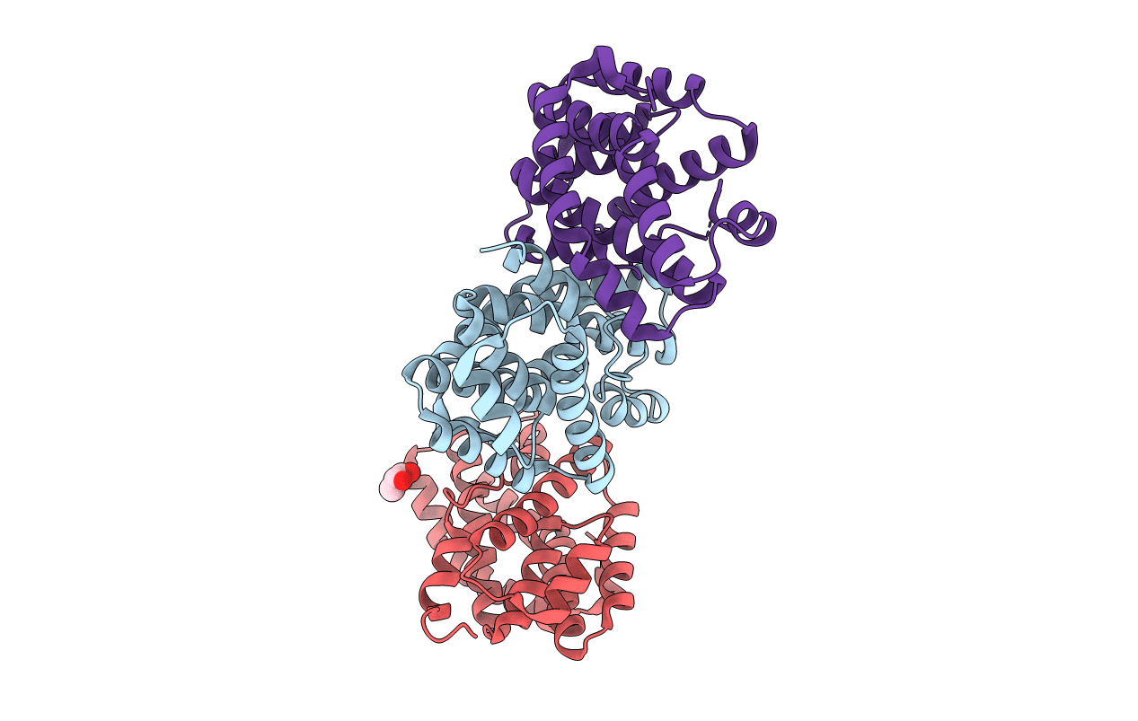
Structure of the Urease Accessory Protein UreF from Helicobacter pylori
Biological Source:
Source Organism:
Host Organism:
PDB Version:
Deposition Date:
2008-04-24
Release Date:
2009-05-12
Method Details:
Experimental Method:
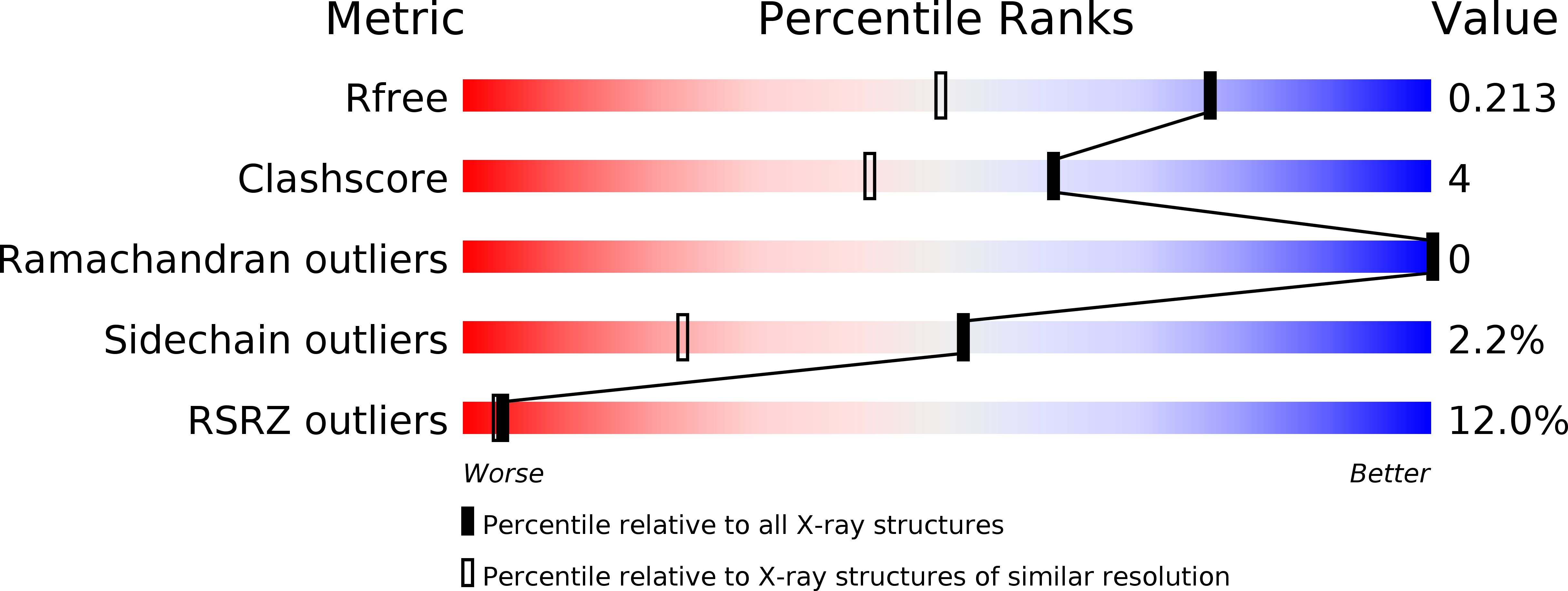
Resolution:
1.55 Å
R-Value Free:
0.21
R-Value Work:
0.18
R-Value Observed:
0.18
Space Group:
C 1 2 1
Macromolecular Entities
Polymer Type:polypeptide(L)
Description:Urease accessory protein ureF
Chain IDs:A, B, C
Chain Length:274
Number of Molecules:3
Biological Source:Helicobacter pylori
Modified Residue
Compound ID Chain ID Parent Comp ID Details 2D Image
MSE A MET SELENOMETHIONINE
Ligand Molecules
Primary Citation
Crystal structure of a truncated urease accessory protein UreF from Helicobacter pylori.
Proteins 78 2839 2848 (2010)
PMID: 20635345 DOI: 10.1002/prot.22802

Abstact

Urease plays a central role in the pathogenesis of Helicobacter pylori in humans. Maturation of this nickel metalloenzyme in bacteria requires the participation of the accessory proteins UreD (termed UreH in H. pylori), UreF, and UreG, which form sequential complexes with the urease apoprotein as well as UreE, a metallochaperone. Here, we describe the crystal structure of C-terminal truncated UreF from H. pylori (residues 1-233), the first UreF structure to be determined, at 1.55 A resolution using SAD methods. UreF forms a dimer in vitro and adopts an all-helical fold congruent with secondary structure prediction. On the basis of evolutionary conservation analysis, the structure reveals a probable binding surface for interaction with other urease components as well as key conserved residues of potential functional relevance.

Legend

Protein

Chemical

Disease

Primary Citation of related structures