3CXH image
Entry Detail
PDB ID:
3CXH
Keywords:
Title:
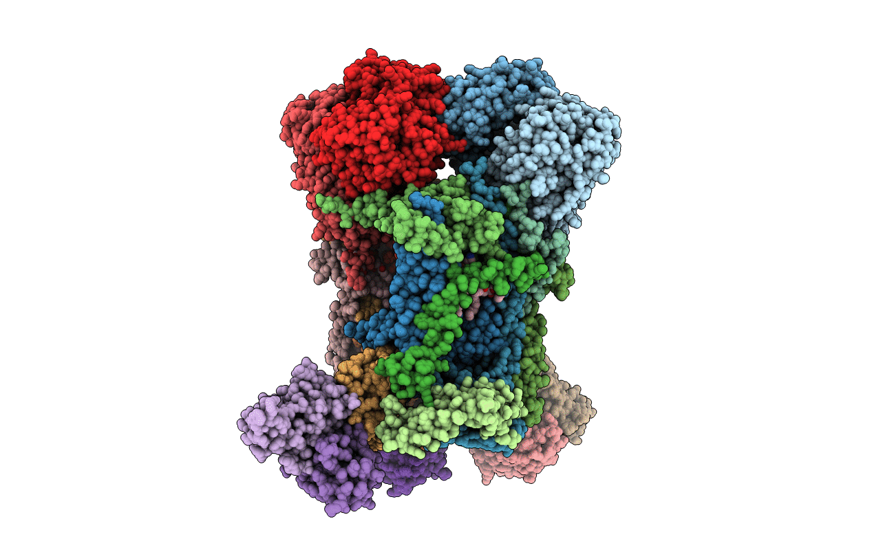
Structure of yeast complex III with isoform-2 cytochrome c bound and definition of a minimal core interface for electron transfer.
Biological Source:
Host Organism:
PDB Version:
Deposition Date:
2008-04-24
Release Date:
2008-05-13
Method Details:
Experimental Method:
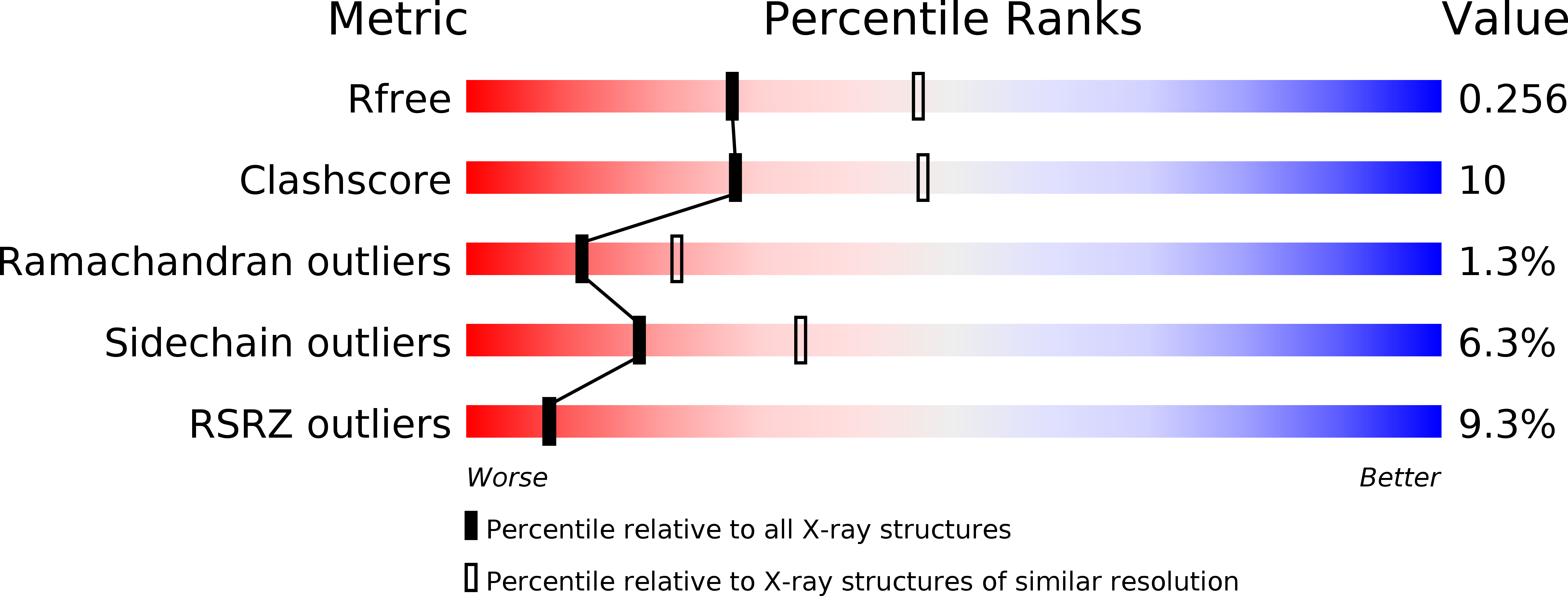
Resolution:
2.50 Å
R-Value Free:
0.25
R-Value Work:
0.22
R-Value Observed:
0.22
Space Group:
P 1 21 1
Macromolecular Entities
Polymer Type:polypeptide(L)
Description:Cytochrome b-c1 complex subunit 1, mitochondrial
Chain IDs:A, L
Chain Length:431
Number of Molecules:2
Biological Source:Saccharomyces cerevisiae
Polymer Type:polypeptide(L)
Description:Cytochrome b-c1 complex subunit 2, mitochondrial
Chain IDs:B, M
Chain Length:352
Number of Molecules:2
Biological Source:Saccharomyces cerevisiae
Polymer Type:polypeptide(L)
Description:Cytochrome b
Chain IDs:C, N
Chain Length:385
Number of Molecules:2
Biological Source:Saccharomyces cerevisiae
Polymer Type:polypeptide(L)
Description:Cytochrome c1, heme protein, mitochondrial
Chain IDs:D, O
Chain Length:248
Number of Molecules:2
Biological Source:Saccharomyces cerevisiae
Polymer Type:polypeptide(L)
Description:Cytochrome b-c1 complex subunit Rieske, mitochondrial
Chain IDs:E, P
Chain Length:185
Number of Molecules:2
Biological Source:Saccharomyces cerevisiae
Polymer Type:polypeptide(L)
Description:Cytochrome b-c1 complex subunit 6
Chain IDs:F, Q
Chain Length:146
Number of Molecules:2
Biological Source:Saccharomyces cerevisiae
Polymer Type:polypeptide(L)
Description:Cytochrome b-c1 complex subunit 7
Chain IDs:G, R
Chain Length:126
Number of Molecules:2
Biological Source:Saccharomyces cerevisiae
Polymer Type:polypeptide(L)
Description:Cytochrome b-c1 complex subunit 8
Chain IDs:H, S
Chain Length:93
Number of Molecules:2
Biological Source:Saccharomyces cerevisiae
Polymer Type:polypeptide(L)
Description:Cytochrome b-c1 complex subunit 9
Chain IDs:I, T
Chain Length:65
Number of Molecules:2
Biological Source:Saccharomyces cerevisiae
Polymer Type:polypeptide(L)
Description:HEAVY CHAIN (VH) OF FV-FRAGMENT
Chain IDs:J, U
Chain Length:127
Number of Molecules:2
Biological Source:Mus musculus
Polymer Type:polypeptide(L)
Description:LIGHT CHAIN (VL) OF FV-FRAGMENT
Chain IDs:K, V
Chain Length:107
Number of Molecules:2
Biological Source:Mus musculus
Polymer Type:polypeptide(L)
Description:Cytochrome c iso-2
Chain IDs:W
Chain Length:112
Number of Molecules:1
Biological Source:Saccharomyces cerevisiae
Modified Residue
Compound ID Chain ID Parent Comp ID Details 2D Image
M3L W LYS N-TRIMETHYLLYSINE
Peptide-like Molecules
PRD_900003
Primary Citation
Structure of complex III with bound cytochrome c in reduced state and definition of a minimal core interface for electron transfer.
J.Biol.Chem. 283 17542 17549 (2008)
PMID: 18390544 DOI: 10.1074/jbc.M710126200

Abstact

In cellular respiration, cytochrome c transfers electrons from cytochrome bc(1) complex (complex III) to cytochrome c oxidase by transiently binding to the membrane proteins. Here, we report the structure of isoform-1 cytochrome c bound to cytochrome bc(1) complex at 1.9 A resolution in reduced state. The dimer structure is asymmetric. Monovalent cytochrome c binding is correlated with conformational changes of the Rieske head domain and subunit QCR6p and with a higher number of interfacial water molecules bound to cytochrome c(1). Pronounced hydration and a "mobility mismatch" at the interface with disordered charged residues on the cytochrome c side are favorable for transient binding. Within the hydrophobic interface, a minimal core was identified by comparison with the novel structure of the complex with bound isoform-2 cytochrome c. Four core interactions encircle the heme cofactors surrounded by variable interactions. The core interface may be a feature to gain specificity for formation of the reactive complex.

Legend

Protein

Chemical

Disease

Primary Citation of related structures