3CX9 image
Deposition Date 2008-04-24
Release Date 2009-04-28
Last Version Date 2024-11-13
Entry Detail
PDB ID:
3CX9
Title:
Crystal Structure of Human serum albumin complexed with Myristic acid and lysophosphatidylethanolamine
Biological Source:
Source Organism:
Homo sapiens (Taxon ID: 9606)
Method Details:
Experimental Method:
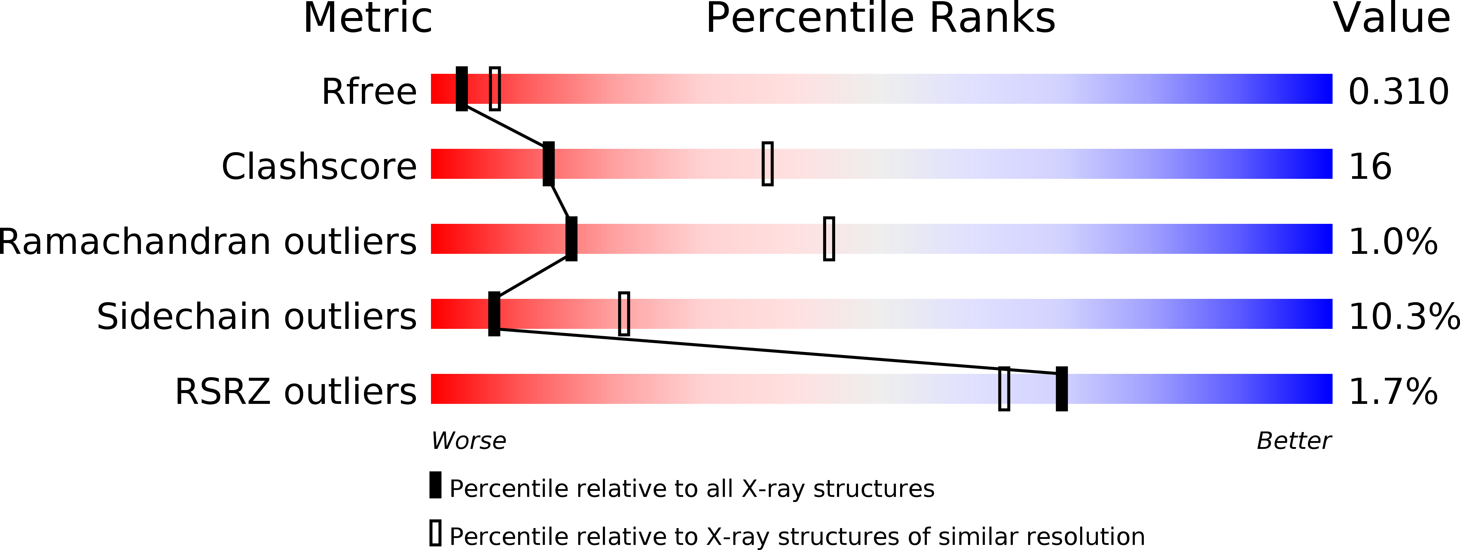
Resolution:
2.80 Å
R-Value Free:
0.29
R-Value Work:
0.21
R-Value Observed:
0.22
Space Group:
C 1 2 1
Macromolecular Entities
Structures with similar UniProt ID
Protein Blast
Polymer Type:polypeptide(L)
Molecule:Serum albumin
Gene (Uniprot):ALB
Chain IDs:A
Chain Length:582
Number of Molecules:1
Biological Source:Homo sapiens
Primary Citation
Structural basis of transport of lysophospholipids by human serum albumin.
Biochem.J. 423 23 30 (2009)
PMID: 19601929 DOI: 10.1042/BJ20090913

Abstact

Lysophospholipids play important roles in cellular signal transduction and are implicated in many biological processes, including tumorigenesis, angiogenesis, immunity, atherosclerosis, arteriosclerosis, cancer and neuronal survival. The intracellular transport of lysophospholipids is through FA (fatty acid)-binding protein. Lysophospholipids are also found in the extracellular space. However, the transport mechanism of lysophospholipids in the extracellular space is unknown. HSA (human serum albumin) is the most abundant carrier protein in blood plasma and plays an important role in determining the absorption, distribution, metabolism and excretion of drugs. In the present study, LPE (lysophosphatidylethanolamine) was used as the ligand to analyse the interaction of lysophospholipids with HSA by fluorescence quenching and crystallography. Fluorescence measurement showed that LPE binds to HSA with a Kd (dissociation constant) of 5.6 microM. The presence of FA (myristate) decreases this binding affinity (Kd of 12.9 microM). Moreover, we determined the crystal structure of HSA in complex with both myristate and LPE and showed that LPE binds at Sudlow site I located in subdomain IIA. LPE occupies two of the three subsites in Sudlow site I, with the LPE acyl chain occupying the hydrophobic bottom of Sudlow site I and the polar head group located at Sudlow site I entrance region pointing to the solvent. This orientation of LPE in HSA suggests that HSA is capable of accommodating other lysophospholipids and phospholipids. The study provides structural information on HSA-lysophospholipid interaction and may facilitate our understanding of the transport and distribution of lysophospholipids.

Legend

Protein

Chemical

Disease

Primary Citation of related structures