Deposition Date
2008-04-23
Release Date
2008-07-01
Last Version Date
2023-08-30
Entry Detail
PDB ID:
3CX2
Keywords:
Title:
Crystal structure of the C1 domain of cardiac isoform of myosin binding protein-C at 1.3A
Biological Source:
Source Organism(s):
Homo sapiens (Taxon ID: 9606)
Expression System(s):
Method Details:
Experimental Method:
Resolution:
1.30 Å
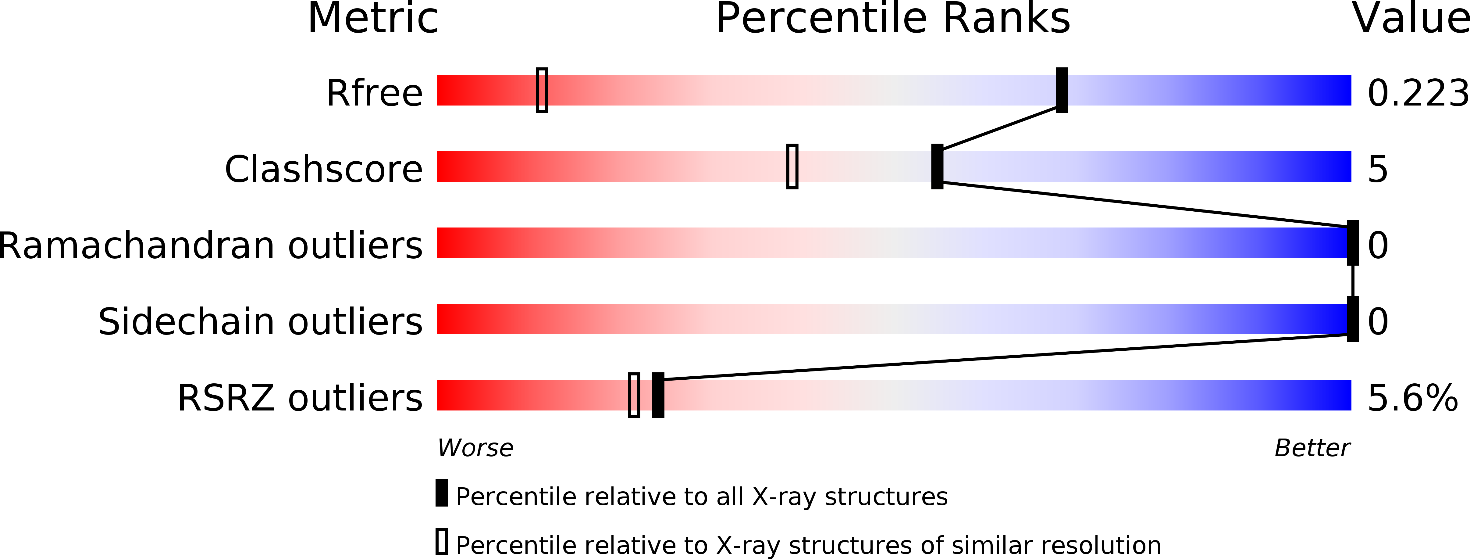
R-Value Free:
0.20
R-Value Observed:
0.16
Space Group:
I 41