Deposition Date
2008-04-21
Release Date
2008-07-01
Last Version Date
2023-08-30
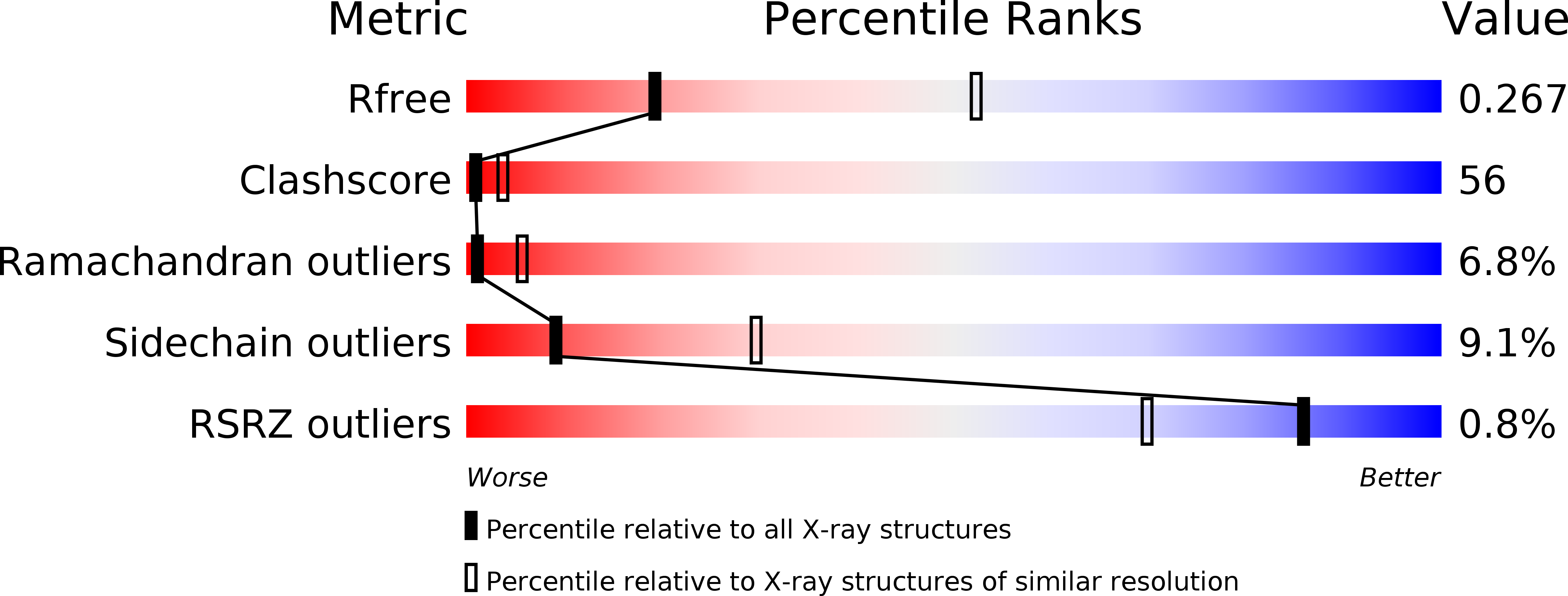
Method Details:
Experimental Method:
Resolution:
3.05 Å
R-Value Free:
0.26
R-Value Work:
0.24
Space Group:
P 31 2 1