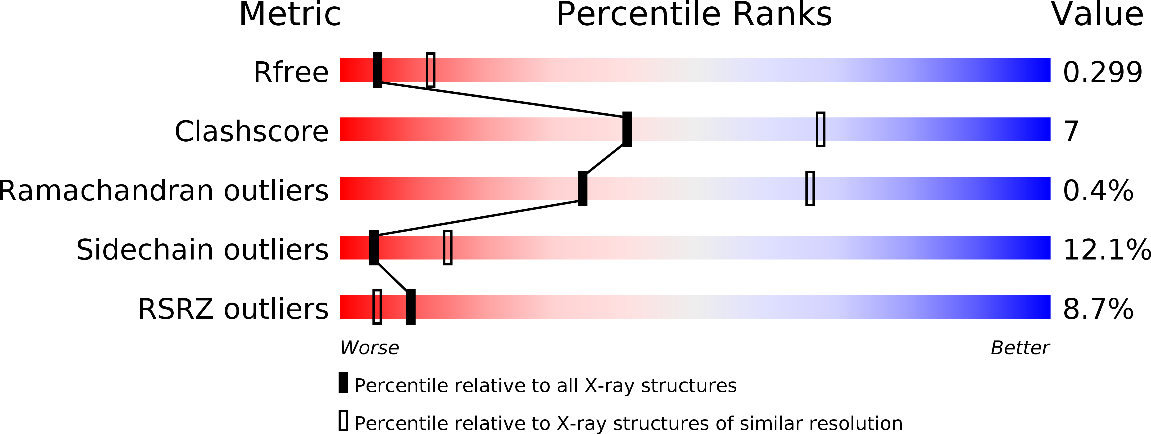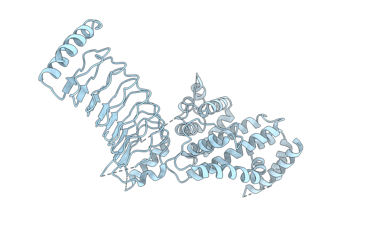
3CVR image
Deposition Date 2008-04-19
Release Date 2008-11-11
Last Version Date 2024-10-30
Entry Detail
PDB ID:
3CVR
Keywords:
Title:
Crystal structure of the full length IpaH3
Biological Source:
Source Organism:
Host Organism:
Method Details:
Experimental Method:
Resolution:
2.80 Å
R-Value Free:
0.27
R-Value Work:
0.25
R-Value Observed:
0.25
Space Group:
P 41 21 2
Macromolecular Entities
Polymer Type:polypeptide(L)
Molecule:Invasion plasmid antigen
Gene (Uniprot):ipaH3
Chain IDs:A
Chain Length:571
Number of Molecules:1
Biological Source:Shigella flexneri 2a
Modified Residue
Compound ID Chain ID Parent Comp ID Details 2D Image
MSE A MET SELENOMETHIONINE
Primary Citation
Structure of a Shigella effector reveals a new class of ubiquitin ligases
Nat.Struct.Mol.Biol. 15 1302 1308 (2008)
PMID: 18997779 DOI: 10.1038/nsmb.1517

Abstact

Bacterial pathogens have evolved effector proteins with ubiquitin E3 ligase activities through structural mimicking. Here we report the crystal structure of the Shigella flexneri type III effector IpaH3, a member of the leucine-rich repeat (LRR)-containing bacterial E3 family. The LRR domain is structurally similar to Yersinia pestis YopM and potentially binds to substrates. The structure of the C-terminal E3 domain differs from the typical RING- and HECT-type E3s. IpaH3 synthesizes a Lys48-linked ubiquitin chain, and the reaction requires noncovalent binding between ubiquitin and a specific E2, UbcH5. Free ubiquitin serves as an acceptor for IpaH3-catalyzed ubiquitin transfer. Cys363 within a conserved CXD motif acts as a nucleophile to catalyze ubiquitin transfer through a transthiolation reaction. The D365N mutant is devoid of E3 activities but turns into a potent ubiquitin-E2 thioesterase. Our analysis establishes a structurally and mechanistically distinct class of ubiquitin ligases found exclusively in pathogenic or symbiotic bacteria.

Legend

Protein

Chemical

Disease

Primary Citation of related structures