3CVI image
Deposition Date 2008-04-18
Release Date 2008-09-23
Last Version Date 2024-10-16
Entry Detail
PDB ID:
3CVI
Keywords:
Title:
How TCR-like antibody recognizes MHC-bound peptide
Biological Source:
Source Organism:
Mus musculus (Taxon ID: 10090)
Method Details:
Experimental Method:
Resolution:
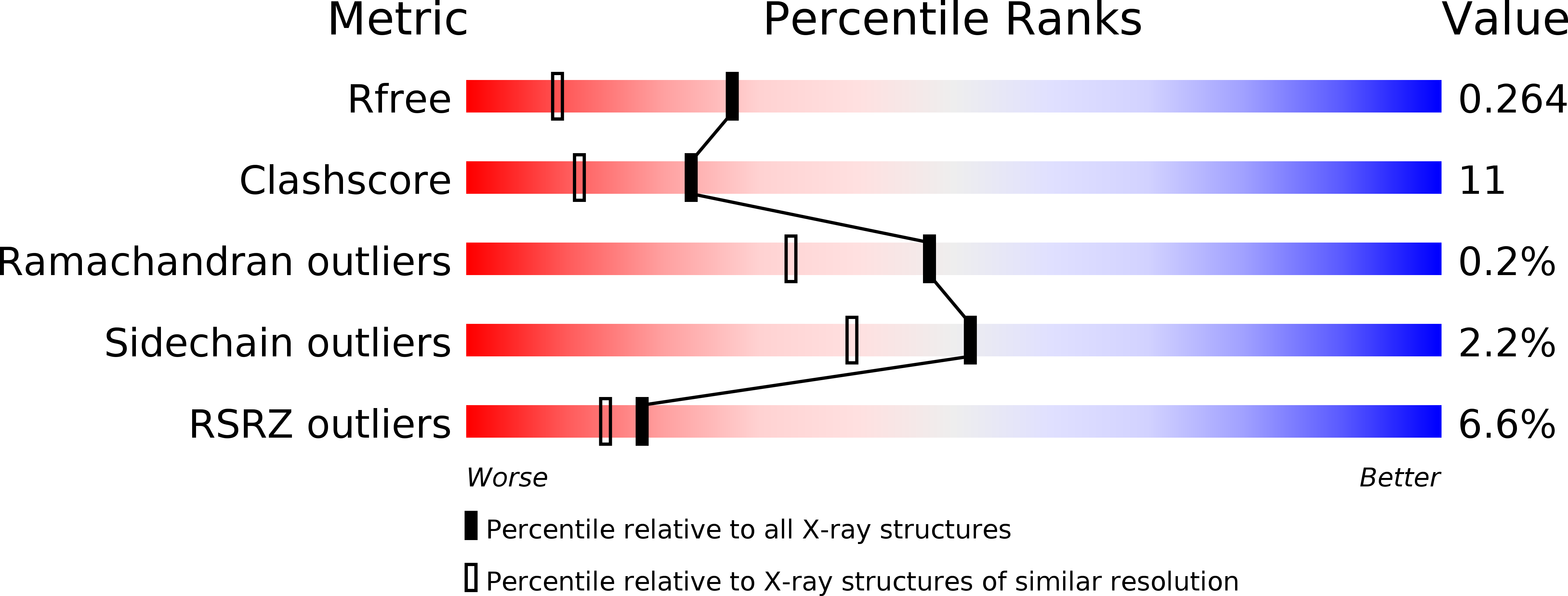
1.80 Å
R-Value Free:
0.26
R-Value Work:
0.21
Space Group:
P 1 21 1
Macromolecular Entities
Polymer Type:polypeptide(L)
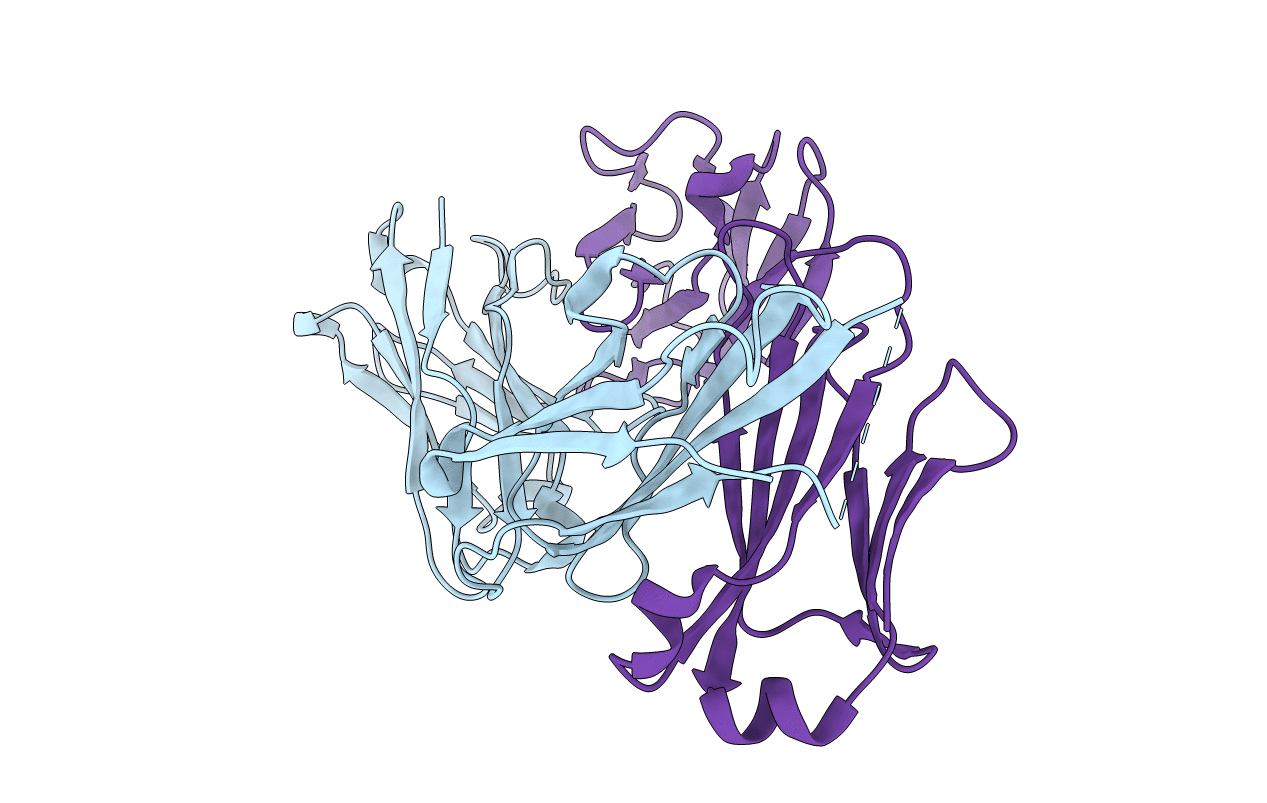
Molecule:25-D1.16 Heavy chain
Chain IDs:A (auth: H)
Chain Length:219
Number of Molecules:1
Biological Source:Mus musculus
Structures with similar UniProt ID
Protein Blast
Polymer Type:polypeptide(L)
Molecule:25-D1.16 Light chain
Chain IDs:B (auth: L)
Chain Length:209
Number of Molecules:1
Biological Source:Mus musculus
Primary Citation
How a T cell receptor-like antibody recognizes major histocompatibility complex-bound peptide
J.Biol.Chem. 283 29053 29059 (2008)
PMID: 18703505 DOI: 10.1074/jbc.M804996200

Abstact

We determined the crystal structures of the T cell receptor (TCR)-like antibody 25-D1.16 Fab fragment bound to a complex of SIINFEKL peptide from ovalbumin and the H-2K(b) molecule. Remarkably, this antibody directly "reads" the structure of the major histocompatibility complex (MHC)-bound peptide, employing the canonical diagonal binding mode utilized by most TCRs. This is in marked contrast with another TCR-like antibody, Hyb3, bound to melanoma peptide MAGE-A1 in association with HLA-A1 MHC class I. Hyb3 assumes a non-canonical orientation over its cognate peptide-MHC and appears to recognize a conformational epitope in which the MHC contribution is dominant. We conclude that TCR-like antibodies can recognize MHC-bound peptide via two different mechanisms: one is similar to that exploited by the preponderance of TCRs and the other requires a non-canonical antibody orientation over the peptide-MHC complex.

Legend

Protein

Chemical

Disease

Primary Citation of related structures